正在加载图片...
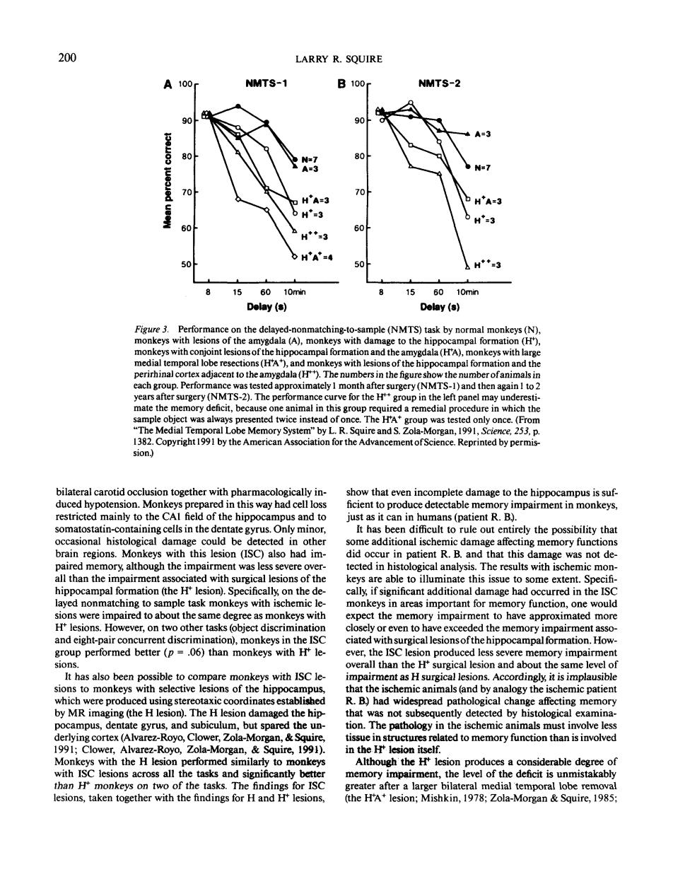
200 LARRY R.SQUIRE A100 NMTS-1 B100 NMTS-2 A= 3 0 10m Delay (s Delay (s) Per (NMTS) ask h N s with h ent to th ala(H) h the fir the num (NMTS-2).The perfo e left pane may und was alw The HA Copyright 1991 by the American on for the Advancement of Sci ence.Reprinted by permis ca duced hy tid on Mor cted ainly to the CA field of the hippocam sional histologica damag coul be ete ted in the e additional isct a ffe ry fund thi na that this da was not de alth han the mpair at ed wi surgical esion of the able to illuminate this issue to some extent ed non to atch with ischem inareas important for mer mpaired to monkeys wit ation).monkeys in 06 all tha the H t as H surgi ns.Ac rdingly,it is impla vere produ using stere oordi ate establishe R B)had wide cha affecting memor and bi the un as not er,Zola gan, lated to memory function than is involvec M ith the performed nila to r gh the H lesion produ a co nsiderable deg the tas after a large ial lesions,taken together with the the H? lesion;Mi shkin,1978:Zola-Morgan Squire,1985 200 A 100 r 90 0 80 1 > S. 70 I so 50 LARRY R. SQUIRE NMTS-1 B I00r 90 80 70 60 50 NMTS-2 A=3 15 60 10nwi Delay (s) 15 60 10min Delay (s) Figure 3. Performance on the delayed-nonmatching-to-sample (NMTS) task by normal monkeys (N), monkeys with lesions of the amygdala (A), monkeys with damage to the hippocampal formation (H*), monkeys with conjoint lesions of the hippocampal formation and the amygdala (H*A), monkeys with large medial temporal lobe resections (H*A+ ), and monkeys with lesions of the hippocampal formation and the perirhinal cortex adjacent to the amygdala (H**). The numbers in the figure show the number of animals in each group. Performance was tested approximately 1 month after surgery (NMTS-1) and then again 1 to 2 years after surgery (NMTS-2). The performance curve for the H*+ group in the left panel may underestimate the memory deficit, because one animal in this group required a remedial procedure in which the sample object was always presented twice instead of once. The WA* group was tested only once. (From "The Medial Temporal Lobe Memory System" by L. R. Squire and S. Zola-Morgan, 1991, Science, 253, p. 1382. Copyright 1991 by the American Association for the Advancement of Science. Reprinted by permission.) bilateral carotid occlusion together with pharmacologically induced hypotension. Monkeys prepared in this way had cell loss restricted mainly to the CA1 field of the hippocampus and to somatostatin-containing cells in the dentate gyrus. Only minor, occasional histological damage could be detected in other brain regions. Monkeys with this lesion (ISC) also had impaired memory, although the impairment was less severe overall than the impairment associated with surgical lesions of the hippocampal formation (the H+ lesion). Specifically, on the delayed nonmatching to sample task monkeys with ischemic lesions were impaired to about the same degree as monkeys with H + lesions. However, on two other tasks (object discrimination and eight-pair concurrent discrimination), monkeys in the ISC group performed better (p = .06) than monkeys with H* lesions. It has also been possible to compare monkeys with ISC lesions to monkeys with selective lesions of the hippocampus, which were produced using stereotaxic coordinates established by MR imaging (the H lesion). The H lesion damaged the hippocampus, dentate gyrus, and subiculum, but spared the underlying cortex (Alvarez-Royo, Glower, Zola-Morgan, & Squire, 1991; Glower, Alvarez-Royo, Zola-Morgan, & Squire, 1991). Monkeys with the H lesion performed similarly to monkeys with ISC lesions across all the tasks and significantly better than JH* monkeys on two of the tasks. The findings for ISC lesions, taken together with the findings for H and H* lesions, show that even incomplete damage to the hippocampus is sufficient to produce detectable memory impairment in monkeys, just as it can in humans (patient R. B.). It has been difficult to rule out entirely the possibility that some additional ischemic damage affecting memory functions did occur in patient R. B. and that this damage was not detected in histological analysis. The results with ischemic monkeys are able to illuminate this issue to some extent. Specifically, if significant additional damage had occurred in the ISC monkeys in areas important for memory function, one would expect the memory impairment to have approximated more closely or even to have exceeded the memory impairment associated with surgical lesions of the hippocampal formation. However, the ISC lesion produced less severe memory impairment overall than the H+ surgical lesion and about the same level of impairment as H surgical lesions. Accordingly, it is implausible that the ischemic animals (and by analogy the ischemic patient R. B.) had widespread pathological change affecting memory that was not subsequently detected by histological examination. The pathology in the ischemic animals must involve less tissue in structures related to memory function than is involved in the № lesion itself. Although the H* lesion produces a considerable degree of memory impairment, the level of the deficit is unmistakably greater after a larger bilateral medial temporal lobe removal (the H+A + lesion; Mishkin, 1978; Zola-Morgan & Squire, 1985;