正在加载图片...
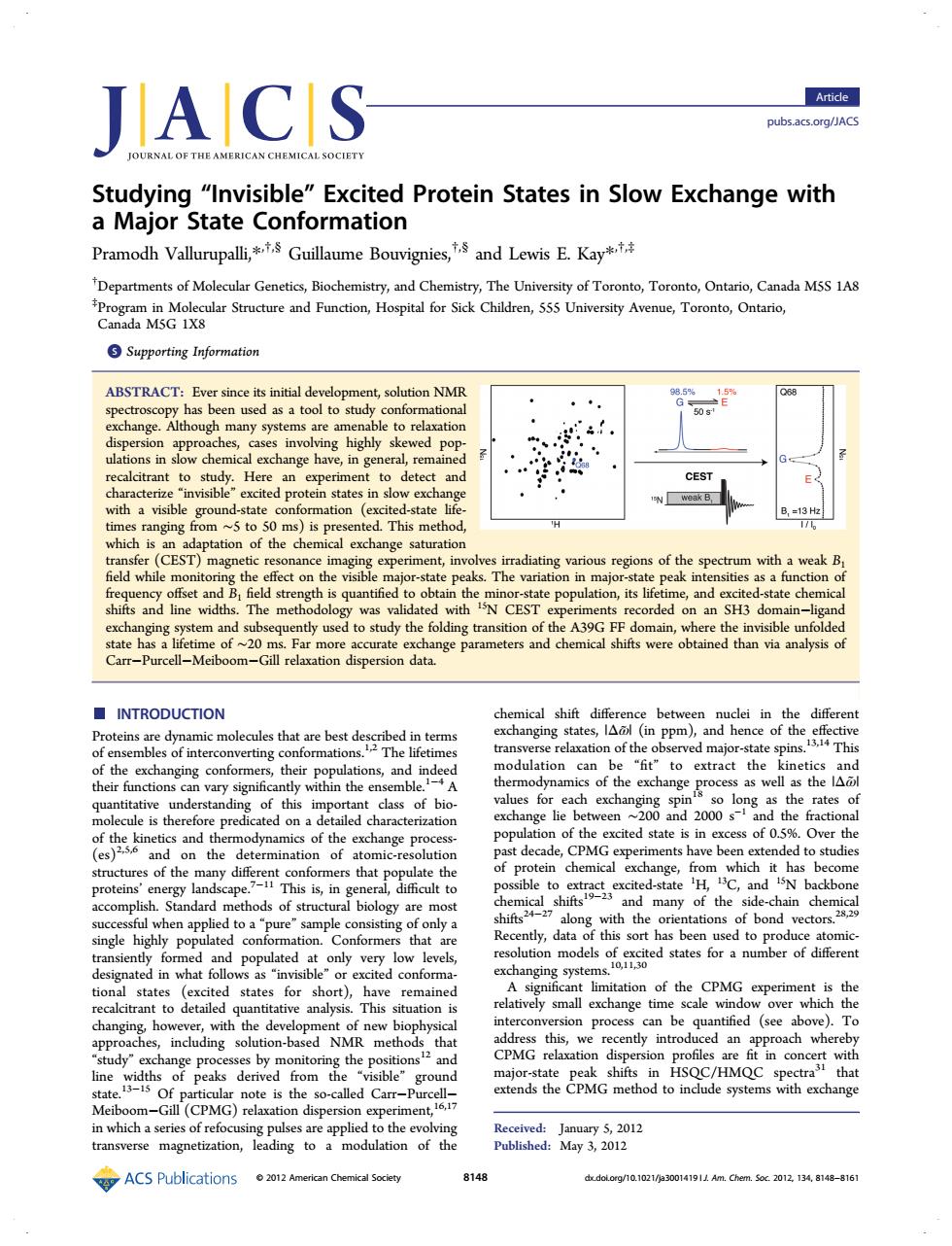
IACS Article pubsacs.org/JACS Studying "Invisible"Excited Protein States in Slow Exchange with a Major State Conformation Pramodh Vallurupalli,Guillaume Bouvigniesand Lewis E.Kay Departments of Molecular Genetics,Biochemistry,and Chemistry,The University of Toronto,Toronto,Ontario,Canada M5S 1A8 nd ion HoptrSik Chlr555vTor Supporting Information ABSTRACT:Ever ce its initial deve re lditranttost He CEST with a visible ground weak B ation rradiating various regions of the spectrum with a weak and ex tate che and tl used to study the foldi of the A39G FF domain,where th and chemical shifts were obtained than via analysis of I-Meil ■INTRODUCTION te can n of the o to cs of the exc s th of this in dlass of bio lie h xchange pro CPM of aton of th chemical exch: ere This s in ge nge, hich ha rotein and many of the side-chain chen ed to apu logy ae vector highly pop t ed states for a number in what very ation of the CPMG is the ge t 6。 d an oach whereb PP the i6ons HSOC om Me (CPMC) with exchange ACS Publications 02012A 8148 d do 10t02 Ldem.5.201213A1-816 Studying “Invisible” Excited Protein States in Slow Exchange with a Major State Conformation Pramodh Vallurupalli,*,†,§ Guillaume Bouvignies,†,§ and Lewis E. Kay*,†,‡ † Departments of Molecular Genetics, Biochemistry, and Chemistry, The University of Toronto, Toronto, Ontario, Canada M5S 1A8 ‡ Program in Molecular Structure and Function, Hospital for Sick Children, 555 University Avenue, Toronto, Ontario, Canada M5G 1X8 *S Supporting Information ABSTRACT: Ever since its initial development, solution NMR spectroscopy has been used as a tool to study conformational exchange. Although many systems are amenable to relaxation dispersion approaches, cases involving highly skewed populations in slow chemical exchange have, in general, remained recalcitrant to study. Here an experiment to detect and characterize “invisible” excited protein states in slow exchange with a visible ground-state conformation (excited-state lifetimes ranging from ∼5 to 50 ms) is presented. This method, which is an adaptation of the chemical exchange saturation transfer (CEST) magnetic resonance imaging experiment, involves irradiating various regions of the spectrum with a weak B1 field while monitoring the effect on the visible major-state peaks. The variation in major-state peak intensities as a function of frequency offset and B1 field strength is quantified to obtain the minor-state population, its lifetime, and excited-state chemical shifts and line widths. The methodology was validated with 15N CEST experiments recorded on an SH3 domain−ligand exchanging system and subsequently used to study the folding transition of the A39G FF domain, where the invisible unfolded state has a lifetime of ∼20 ms. Far more accurate exchange parameters and chemical shifts were obtained than via analysis of Carr−Purcell−Meiboom−Gill relaxation dispersion data. ■ INTRODUCTION Proteins are dynamic molecules that are best described in terms of ensembles of interconverting conformations.1,2 The lifetimes of the exchanging conformers, their populations, and indeed their functions can vary significantly within the ensemble.1−4 A quantitative understanding of this important class of biomolecule is therefore predicated on a detailed characterization of the kinetics and thermodynamics of the exchange process- (es)2,5,6 and on the determination of atomic-resolution structures of the many different conformers that populate the proteins’ energy landscape.7−11 This is, in general, difficult to accomplish. Standard methods of structural biology are most successful when applied to a “pure” sample consisting of only a single highly populated conformation. Conformers that are transiently formed and populated at only very low levels, designated in what follows as “invisible” or excited conformational states (excited states for short), have remained recalcitrant to detailed quantitative analysis. This situation is changing, however, with the development of new biophysical approaches, including solution-based NMR methods that “study” exchange processes by monitoring the positions12 and line widths of peaks derived from the “visible” ground state.13−15 Of particular note is the so-called Carr−Purcell− Meiboom−Gill (CPMG) relaxation dispersion experiment,16,17 in which a series of refocusing pulses are applied to the evolving transverse magnetization, leading to a modulation of the chemical shift difference between nuclei in the different exchanging states, |Δω̃| (in ppm), and hence of the effective transverse relaxation of the observed major-state spins.13,14 This modulation can be “fit” to extract the kinetics and thermodynamics of the exchange process as well as the |Δω̃| values for each exchanging spin18 so long as the rates of exchange lie between ∼200 and 2000 s−1 and the fractional population of the excited state is in excess of 0.5%. Over the past decade, CPMG experiments have been extended to studies of protein chemical exchange, from which it has become possible to extract excited-state 1 H, 13C, and 15N backbone chemical shifts19−23 and many of the side-chain chemical shifts24−27 along with the orientations of bond vectors.28,29 Recently, data of this sort has been used to produce atomicresolution models of excited states for a number of different exchanging systems.10,11,30 A significant limitation of the CPMG experiment is the relatively small exchange time scale window over which the interconversion process can be quantified (see above). To address this, we recently introduced an approach whereby CPMG relaxation dispersion profiles are fit in concert with major-state peak shifts in HSQC/HMQC spectra31 that extends the CPMG method to include systems with exchange Received: January 5, 2012 Published: May 3, 2012 Article pubs.acs.org/JACS © 2012 American Chemical Society 8148 dx.doi.org/10.1021/ja3001419 | J. Am. Chem. Soc. 2012, 134, 8148−8161