正在加载图片...

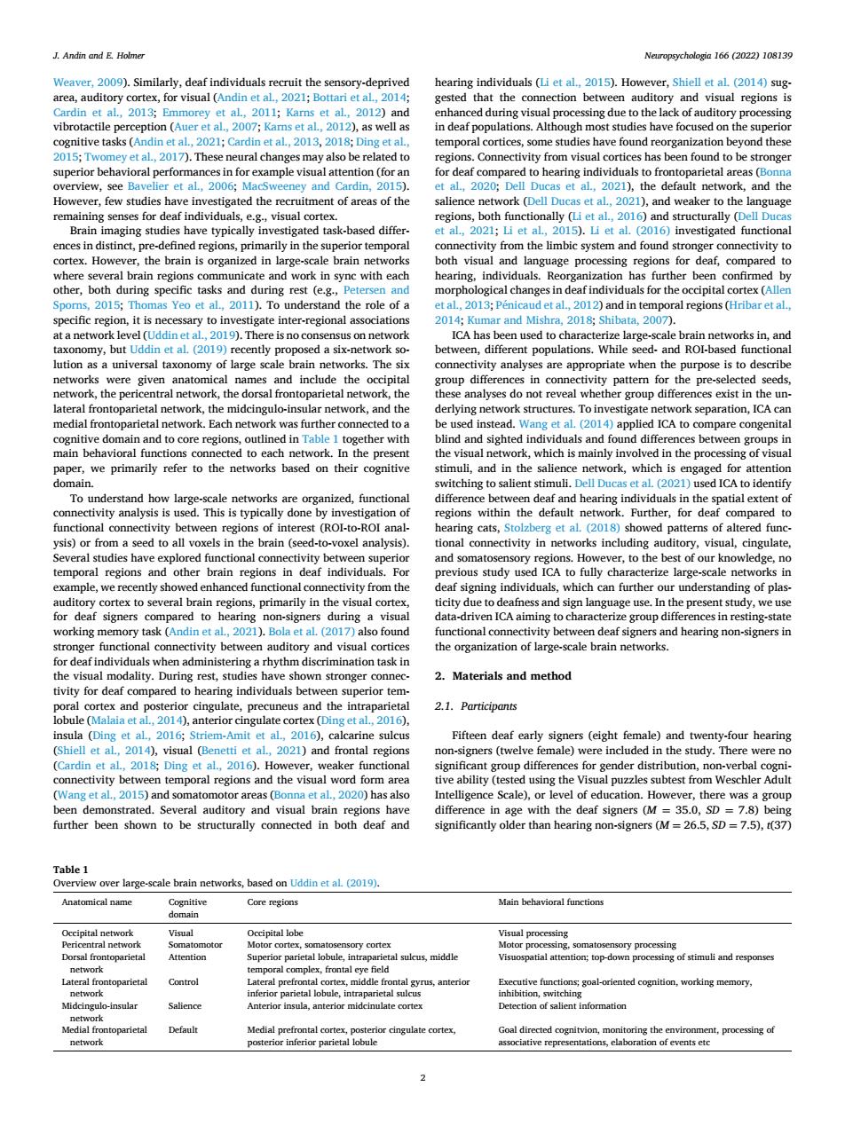
J.Andin and E Holmer mp chologin166(2022)10813 Ding et al as ork (Dell Duc t al.2 nd weaker to the lan eeriortempor ctivityto urther s Ye d the role of a 2012) poralegonsGHri e)m on ne ICA has 2018sS ks in network,a ork separation,ICA ca ether s bet paper we primarily refer to the networks based on their cognitive u,and in the salience ne ork.which is ale and h ring d.This is typi with he defa brain (seed voxel analy ctivity in orks includ erize larg x to eral brain region s,prim ly in the visual ue to eafness and sign langu euse.I e present study nal co ivity betw he organization ot large.scale brain network 2.Materials and method 2.1.Participants insula (Ding et al 2016 Amit et al. Fifteen deaf early signers(eight female)and tw enty-four hearing ere n et a 2018:Dinz et al 2016).H to be Table 1 works,based on ictaNeuropsychologia 166 (2022) 108139 2 Weaver, 2009). Similarly, deaf individuals recruit the sensory-deprived area, auditory cortex, for visual (Andin et al., 2021; Bottari et al., 2014; Cardin et al., 2013; Emmorey et al., 2011; Karns et al., 2012) and vibrotactile perception (Auer et al., 2007; Karns et al., 2012), as well as cognitive tasks (Andin et al., 2021; Cardin et al., 2013, 2018; Ding et al., 2015; Twomey et al., 2017). These neural changes may also be related to superior behavioral performances in for example visual attention (for an overview, see Bavelier et al., 2006; MacSweeney and Cardin, 2015). However, few studies have investigated the recruitment of areas of the remaining senses for deaf individuals, e.g., visual cortex. Brain imaging studies have typically investigated task-based differences in distinct, pre-defined regions, primarily in the superior temporal cortex. However, the brain is organized in large-scale brain networks where several brain regions communicate and work in sync with each other, both during specific tasks and during rest (e.g., Petersen and Sporns, 2015; Thomas Yeo et al., 2011). To understand the role of a specific region, it is necessary to investigate inter-regional associations at a network level (Uddin et al., 2019). There is no consensus on network taxonomy, but Uddin et al. (2019) recently proposed a six-network solution as a universal taxonomy of large scale brain networks. The six networks were given anatomical names and include the occipital network, the pericentral network, the dorsal frontoparietal network, the lateral frontoparietal network, the midcingulo-insular network, and the medial frontoparietal network. Each network was further connected to a cognitive domain and to core regions, outlined in Table 1 together with main behavioral functions connected to each network. In the present paper, we primarily refer to the networks based on their cognitive domain. To understand how large-scale networks are organized, functional connectivity analysis is used. This is typically done by investigation of functional connectivity between regions of interest (ROI-to-ROI analysis) or from a seed to all voxels in the brain (seed-to-voxel analysis). Several studies have explored functional connectivity between superior temporal regions and other brain regions in deaf individuals. For example, we recently showed enhanced functional connectivity from the auditory cortex to several brain regions, primarily in the visual cortex, for deaf signers compared to hearing non-signers during a visual working memory task (Andin et al., 2021). Bola et al. (2017) also found stronger functional connectivity between auditory and visual cortices for deaf individuals when administering a rhythm discrimination task in the visual modality. During rest, studies have shown stronger connectivity for deaf compared to hearing individuals between superior temporal cortex and posterior cingulate, precuneus and the intraparietal lobule (Malaia et al., 2014), anterior cingulate cortex (Ding et al., 2016), insula (Ding et al., 2016; Striem-Amit et al., 2016), calcarine sulcus (Shiell et al., 2014), visual (Benetti et al., 2021) and frontal regions (Cardin et al., 2018; Ding et al., 2016). However, weaker functional connectivity between temporal regions and the visual word form area (Wang et al., 2015) and somatomotor areas (Bonna et al., 2020) has also been demonstrated. Several auditory and visual brain regions have further been shown to be structurally connected in both deaf and hearing individuals (Li et al., 2015). However, Shiell et al. (2014) suggested that the connection between auditory and visual regions is enhanced during visual processing due to the lack of auditory processing in deaf populations. Although most studies have focused on the superior temporal cortices, some studies have found reorganization beyond these regions. Connectivity from visual cortices has been found to be stronger for deaf compared to hearing individuals to frontoparietal areas (Bonna et al., 2020; Dell Ducas et al., 2021), the default network, and the salience network (Dell Ducas et al., 2021), and weaker to the language regions, both functionally (Li et al., 2016) and structurally (Dell Ducas et al., 2021; Li et al., 2015). Li et al. (2016) investigated functional connectivity from the limbic system and found stronger connectivity to both visual and language processing regions for deaf, compared to hearing, individuals. Reorganization has further been confirmed by morphological changes in deaf individuals for the occipital cortex (Allen et al., 2013; P´enicaud et al., 2012) and in temporal regions (Hribar et al., 2014; Kumar and Mishra, 2018; Shibata, 2007). ICA has been used to characterize large-scale brain networks in, and between, different populations. While seed- and ROI-based functional connectivity analyses are appropriate when the purpose is to describe group differences in connectivity pattern for the pre-selected seeds, these analyses do not reveal whether group differences exist in the underlying network structures. To investigate network separation, ICA can be used instead. Wang et al. (2014) applied ICA to compare congenital blind and sighted individuals and found differences between groups in the visual network, which is mainly involved in the processing of visual stimuli, and in the salience network, which is engaged for attention switching to salient stimuli. Dell Ducas et al. (2021) used ICA to identify difference between deaf and hearing individuals in the spatial extent of regions within the default network. Further, for deaf compared to hearing cats, Stolzberg et al. (2018) showed patterns of altered functional connectivity in networks including auditory, visual, cingulate, and somatosensory regions. However, to the best of our knowledge, no previous study used ICA to fully characterize large-scale networks in deaf signing individuals, which can further our understanding of plasticity due to deafness and sign language use. In the present study, we use data-driven ICA aiming to characterize group differences in resting-state functional connectivity between deaf signers and hearing non-signers in the organization of large-scale brain networks. 2. Materials and method 2.1. Participants Fifteen deaf early signers (eight female) and twenty-four hearing non-signers (twelve female) were included in the study. There were no significant group differences for gender distribution, non-verbal cognitive ability (tested using the Visual puzzles subtest from Weschler Adult Intelligence Scale), or level of education. However, there was a group difference in age with the deaf signers (M = 35.0, SD = 7.8) being significantly older than hearing non-signers (M = 26.5, SD = 7.5), t(37) Table 1 Overview over large-scale brain networks, based on Uddin et al. (2019). Anatomical name Cognitive domain Core regions Main behavioral functions Occipital network Visual Occipital lobe Visual processing Pericentral network Somatomotor Motor cortex, somatosensory cortex Motor processing, somatosensory processing Dorsal frontoparietal network Attention Superior parietal lobule, intraparietal sulcus, middle temporal complex, frontal eye field Visuospatial attention; top-down processing of stimuli and responses Lateral frontoparietal network Control Lateral prefrontal cortex, middle frontal gyrus, anterior inferior parietal lobule, intraparietal sulcus Executive functions; goal-oriented cognition, working memory, inhibition, switching Midcingulo-insular network Salience Anterior insula, anterior midcinulate cortex Detection of salient information Medial frontoparietal network Default Medial prefrontal cortex, posterior cingulate cortex, posterior inferior parietal lobule Goal directed cognitvion, monitoring the environment, processing of associative representations, elaboration of events etc J. Andin and E. Holmer