正在加载图片...
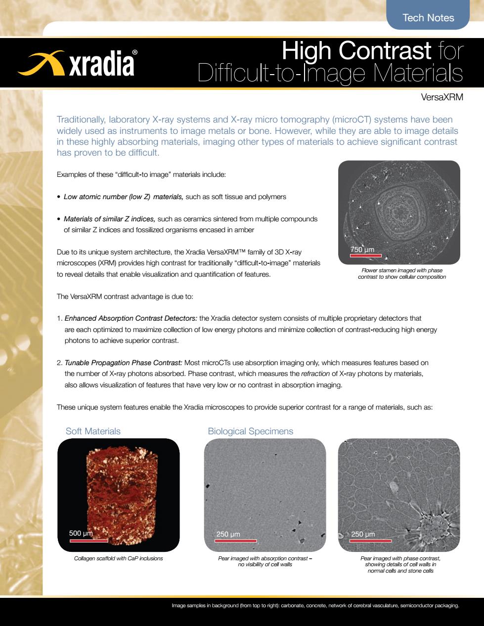
Tech Notes 元xradia High Contrast for Difficult-to-Image Materials VersaXRM Traditionally,laboratory X-ray systems and X-ray micro tomography(microCT)systems have been widely used as instruments to image metals or bone.However,while they are able to image details in these highly absorbing materials,imaging other types of materials to achieve significant contrast has proven to be difficult. Examples of these"difficult-to image"materials include: Low atomic number (low Z)materials,such as soft tissue and polymers Materials of similar Z indices,such as ceramics sintered from multiple compounds of similar Z indices and fossilized organisms encased in amber Due to its unique system architecture,the Xradia VersaXRMTM family of 3D X-ray 750μm2 microscopes(XRM)provides high contrast for traditionally "difficult-to-image"materials to reveal details that enable visualization and quantification of features. Flower stamen imaged with phase contrast to show cellular composition The VersaXRM contrast advantage is due to: 1.Enhanced Absorption Contrast Detectors:the Xradia detector system consists of multiple proprietary detectors that are each optimized to maximize collection of low energy photons and minimize collection of contrast-reducing high energy photons to achieve superior contrast. 2.Tunable Propagation Phase Contrast:Most microCTs use absorption imaging only,which measures features based on the number of X-ray photons absorbed.Phase contrast,which measures the refraction of X-ray photons by materials, also allows visualization of features that have very low or no contrast in absorption imaging. These unique system features enable the Xradia microscopes to provide superior contrast for a range of materials,such as: Soft Materials Biological Specimens 500μm 250μm 250pm Collagen scaffold with CaP inclusions Pear imaged with absorption contrast- Pear imaged with phase contrast, no visibility of cell walls showing details of cell walls in nomal cells and stone cells Image samples in background (from top to right):carbonate,concrete,network of cerebral vasculature,semiconductor packagingHigh Contrast for Difficult-to-Image Materials VersaXRM Tech Notes Traditionally, laboratory X-ray systems and X-ray micro tomography (microCT) systems have been widely used as instruments to image metals or bone. However, while they are able to image details in these highly absorbing materials, imaging other types of materials to achieve significant contrast has proven to be difficult. Examples of these “difficult-to image” materials include: • Low atomic number (low Z) materials, such as soft tissue and polymers • Materials of similar Z indices, such as ceramics sintered from multiple compounds of similar Z indices and fossilized organisms encased in amber Due to its unique system architecture, the Xradia VersaXRM™ family of 3D X-ray microscopes (XRM) provides high contrast for traditionally “difficult-to-image” materials to reveal details that enable visualization and quantification of features. The VersaXRM contrast advantage is due to: 1. Enhanced Absorption Contrast Detectors: the Xradia detector system consists of multiple proprietary detectors that are each optimized to maximize collection of low energy photons and minimize collection of contrast-reducing high energy photons to achieve superior contrast. 2. Tunable Propagation Phase Contrast: Most microCTs use absorption imaging only, which measures features based on the number of X-ray photons absorbed. Phase contrast, which measures the refraction of X-ray photons by materials, also allows visualization of features that have very low or no contrast in absorption imaging. These unique system features enable the Xradia microscopes to provide superior contrast for a range of materials, such as: Soft Materials Biological Specimens Collagen scaffold with CaP inclusions Pear imaged with absorption contrast – no visibility of cell walls Pear imaged with phase contrast, showing details of cell walls in normal cells and stone cells Image samples in background (from top to right): carbonate, concrete, network of cerebral vasculature, semiconductor packaging. Flower stamen imaged with phase contrast to show cellular composition 750 µm 500 µm 250 µm 250 µm