正在加载图片...
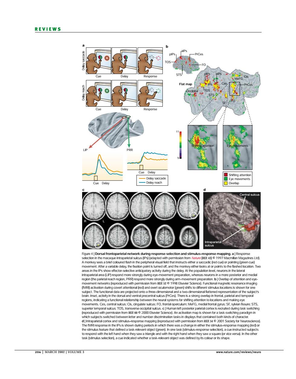
REVIEWS cue n d by its coour or ts shaoe 206 MARCH2002 VOLUM正 206 | MARCH 2002 | VOLUME 3 www.nature.com/reviews/neuro REVIEWS a b c d Cue LIP R L Cue Delay Cue Delay Delay Response Cue Delay Response Delay saccade TOS TOS Ces Cis MeFG Shifting attention Shifting attention Eye movements Eye movements Overlap 11 5 Occipital Flat map STS SF STS FO FO PrCes PrCes aIPs pIPs IPs pIPs aIPs PRR Intraparietal sulcus Central sulcus Delay saccade Delay reach Delay reach Figure 4 | Dorsal frontoparietal network during response selection and stimulus–response mapping. a | Response selection in the macaque intraparietal sulcus (IPs) (adapted with permission from Nature (REF. 43) © 1997 Macmillan Magazines Ltd). A monkey sees a brief coloured flash in the peripheral visual field that instructs either a saccadic (red cue) or pointing (green cue) movement. After a variable delay, the fixation point is turned off, and the monkey either looks at or points to the flashed location. Two areas in the IPs show effector-selective anticipatory activity during the delay. At the population level, neurons in the lateral intraparietal area (LIP) respond more strongly during eye-movement preparation, whereas neurons in a more posterior and medial region (the parietal reach region, PRR) respond more strongly during arm-movement preparation. b | Overlap of attention and eyemovement networks (reproduced with permission from REF. 52 © 1998 Elsevier Science). Functional magnetic resonance imaging (fMRI) activation during covert attentional (red) and overt oculomotor (green) shifts to different stimulus locations is shown for one subject. The functional data are projected onto a three-dimensional and a two-dimensional flattened representation of the subject’s brain. Inset, activity in the dorsal and ventral precentral sulcus (PrCes). There is a strong overlap in frontal, parietal and temporal regions, indicating a functional relationship between the neural systems for shifting attention to locations and making eye movements. Ces, central sulcus; Cis, cingulate sulcus; FO, frontal operculum; MeFG, medial frontal gyrus; SF, sylvian fissure; STS, superior temporal sulcus; TOS, transverse occipital sulcus. c | Human left posterior parietal cortex is recruited during task switching (reproduced with permission from REF. 60 © 2000 Elsevier Science). An activation map is shown for a task-switching paradigm in which subjects switched between letter and number discrimination tasks in displays that contained both kinds of character. d | Intraparietal cortex and stimulus–response mapping (reproduced with permission from REF. 54 © 2001 Society for Neuroscience). The fMRI response in the IPs is shown during periods in which there was a change in either the stimulus–response mapping (red) or the stimulus feature that defined a task-relevant object (green). In one task (stimulus–response selection), a cue instructed subjects to respond with the left hand when they saw a triangle and with the right hand when they saw a square (or vice versa). In the other task (stimulus selection), a cue indicated whether a task-relevant object was defined by its colour or its shape