正在加载图片...
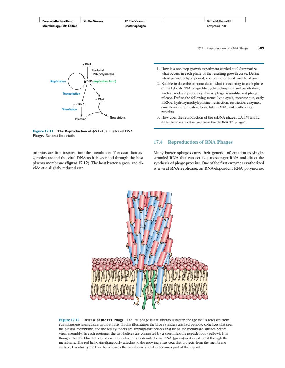
he vire 17.4 Reproduction of RNA Phages 389 he Repoduction ofStrand DN 17.4 Reproduction of RNA Phages es synthesized e17.12 The Pf that is nd the re nhinathic hat lic surface hefo surface.E ally the blue belix leaves the men nd also becomes part of the capsid Prescott−Harley−Klein: Microbiology, Fifth Edition VI. The Viruses 17. The Viruses: Bacteriophages © The McGraw−Hill Companies, 2002 proteins are first inserted into the membrane. The coat then assembles around the viral DNA as it is secreted through the host plasma membrane (figure 17.12). The host bacteria grow and divide at a slightly reduced rate. 1. How is a one-step growth experiment carried out? Summarize what occurs in each phase of the resulting growth curve. Define latent period, eclipse period, rise period or burst, and burst size. 2. Be able to describe in some detail what is occurring in each phase of the lytic dsDNA phage life cycle: adsorption and penetration, nucleic acid and protein synthesis, phage assembly, and phage release. Define the following terms: lytic cycle, receptor site, early mRNA, hydroxymethylcytosine, restriction, restriction enzymes, concatemers, replicative form, late mRNA, and scaffolding proteins. 3. How does the reproduction of the ssDNA phages X174 and fd differ from each other and from the dsDNA T4 phage? 17.4 Reproduction of RNA Phages Many bacteriophages carry their genetic information as singlestranded RNA that can act as a messenger RNA and direct the synthesis of phage proteins. One of the first enzymes synthesized is a viral RNA replicase, an RNA-dependent RNA polymerase 17.4 Reproduction of RNA Phages 389 Figure 17.12 Release of the Pf1 Phage. The Pf1 phage is a filamentous bacteriophage that is released from Pseudomonas aeruginosa without lysis. In this illustration the blue cylinders are hydrophobic α-helices that span the plasma membrane, and the red cylinders are amphipathic helices that lie on the membrane surface before virus assembly. In each protomer the two helices are connected by a short, flexible peptide loop (yellow). It is thought that the blue helix binds with circular, single-stranded viral DNA (green) as it is extruded through the membrane. The red helix simultaneously attaches to the growing virus coat that projects from the membrane surface. Eventually the blue helix leaves the membrane and also becomes part of the capsid. Figure 17.11 The Reproduction of X174, a Strand DNA Phage. See text for details. + DNA Bacterial DNA polymerase Replication ± DNA (replicative form) Transcription + mRNA + DNA Proteins New virions Translation�