正在加载图片...
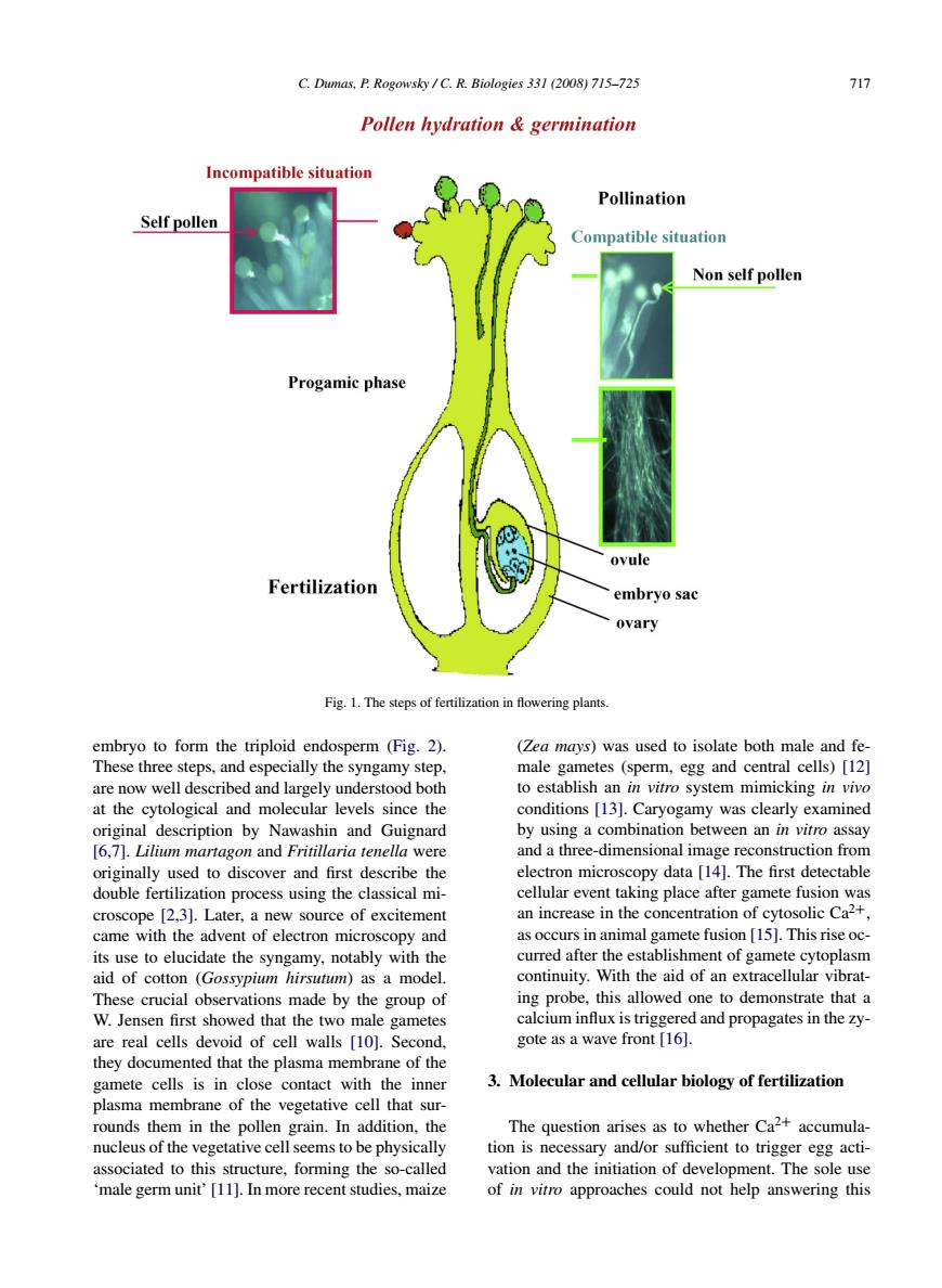
C.Dumas,P Rogowsky/C.R.Biologles 331(2008)715-725 71 Pollen hydration germination Incompatible situation Pollination Self pollen Compatible situation Non self pollen Progamic phase ovule embryo sac ovary Fig1.The steps of fertilization in flowering plants. embryo to form the triploid endosperm(Fig.2). (Zea mays)was used to isolate both male and fe These three steps.and especially the syngamy step. male gametes (sperm,egg and central cells)[12] are now well described and largely understood both to establish an in vitro system mimicking in vivo at the cytological and molecular levels since the conditions [1 3].Caryogamy was clearly examined mb between an in viro assa on n cover and I m cEctonmi using the (2.3).Lat an incr eve in the ad as oceurs in animal o nete fusion [15].This rise oc its use to elucidate the s notably with the curred after the establishment of gamete cytoplasm continuity.With the aid of an extracellular vibrat- These crucial observations made by the group of ing probe,this allowed one to demonstrate that a w Jensen first showed that the two male gametes calcium influx is triggered and propagates in the zy- are real cells devoid of cell walls [10].Second gote as a wave front [16]. they documented that the plasma membrane of the gamete cells is in close contact with the inner 3.Molecular and cellular biology of fertilizatior plasma membrane of the vegetative cell that sur- rounds them in the pollen the The question arises as to whether Caz+accumula tion is necessary and/or su ent to trigger egg act cture,forming the and the initiation of deve The vitro approaches could C. Dumas, P. Rogowsky / C. R. Biologies 331 (2008) 715–725 717 Fig. 1. The steps of fertilization in flowering plants. embryo to form the triploid endosperm (Fig. 2). These three steps, and especially the syngamy step, are now well described and largely understood both at the cytological and molecular levels since the original description by Nawashin and Guignard [6,7]. Lilium martagon and Fritillaria tenella were originally used to discover and first describe the double fertilization process using the classical microscope [2,3]. Later, a new source of excitement came with the advent of electron microscopy and its use to elucidate the syngamy, notably with the aid of cotton (Gossypium hirsutum) as a model. These crucial observations made by the group of W. Jensen first showed that the two male gametes are real cells devoid of cell walls [10]. Second, they documented that the plasma membrane of the gamete cells is in close contact with the inner plasma membrane of the vegetative cell that surrounds them in the pollen grain. In addition, the nucleus of the vegetative cell seems to be physically associated to this structure, forming the so-called ‘male germ unit’ [11]. In more recent studies, maize (Zea mays) was used to isolate both male and female gametes (sperm, egg and central cells) [12] to establish an in vitro system mimicking in vivo conditions [13]. Caryogamy was clearly examined by using a combination between an in vitro assay and a three-dimensional image reconstruction from electron microscopy data [14]. The first detectable cellular event taking place after gamete fusion was an increase in the concentration of cytosolic Ca2+, as occurs in animal gamete fusion [15]. This rise occurred after the establishment of gamete cytoplasm continuity. With the aid of an extracellular vibrating probe, this allowed one to demonstrate that a calcium influx is triggered and propagates in the zygote as a wave front [16]. 3. Molecular and cellular biology of fertilization The question arises as to whether Ca2+ accumulation is necessary and/or sufficient to trigger egg activation and the initiation of development. The sole use of in vitro approaches could not help answering this