正在加载图片...
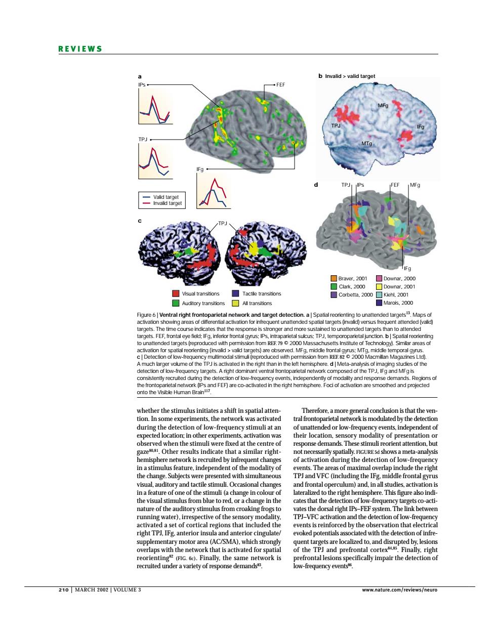
REVIEWS b Invalld>valld targe Eaa lus initia e of los nded or k stimuli ation durine the det ction of l the naf sal right i het sthat included th hat el k d for sp the T atthedeiecg ecruited under a v 210 MARCH2002 VOLUM正3 210 | MARCH 2002 | VOLUME 3 www.nature.com/reviews/neuro REVIEWS Therefore, a more general conclusion is that the ventral frontoparietal network is modulated by the detection of unattended or low-frequency events, independent of their location, sensory modality of presentation or response demands. These stimuli reorient attention, but not necessarily spatially. FIGURE 5d shows a meta-analysis of activation during the detection of low-frequency events. The areas of maximal overlap include the right TPJ and VFC (including the IFg, middle frontal gyrus and frontal operculum) and, in all studies, activation is lateralized to the right hemisphere. This figure also indicates that the detection of low-frequency targets co-activates the dorsal right IPs–FEF system. The link between TPJ–VFC activation and the detection of low-frequency events is reinforced by the observation that electrical evoked potentials associated with the detection of infrequent targets are localized to, and disrupted by, lesions of the TPJ and prefrontal cortex84,85. Finally, right prefrontal lesions specifically impair the detection of low-frequency events86. whether the stimulus initiates a shift in spatial attention. In some experiments, the network was activated during the detection of low-frequency stimuli at an expected location; in other experiments, activation was observed when the stimuli were fixed at the centre of gaze80,81. Other results indicate that a similar righthemisphere network is recruited by infrequent changes in a stimulus feature, independent of the modality of the change. Subjects were presented with simultaneous visual, auditory and tactile stimuli. Occasional changes in a feature of one of the stimuli (a change in colour of the visual stimulus from blue to red, or a change in the nature of the auditory stimulus from croaking frogs to running water), irrespective of the sensory modality, activated a set of cortical regions that included the right TPJ, IFg, anterior insula and anterior cingulate/ supplementary motor area (AC/SMA), which strongly overlaps with the network that is activated for spatial reorienting82 (FIG. 6c). Finally, the same network is recruited under a variety of response demands83. b Invalid > valid target IPs TPJ IFg FEF TPJ a d c Valid target Invalid target IFg MFg MTg TPJ Visual transitions Auditory transitions All transitions Braver, 2001 Downar, 2000 Clark, 2000 Downar, 2001 Corbetta, 2000 Kiehl, 2001 Marois, 2000 Tactile transitions TPJ MFg IPs FEF IFg Figure 6 | Ventral right frontoparietal network and target detection. a | Spatial reorienting to unattended targets13. Maps of activation showing areas of differential activation for infrequent unattended spatial targets (invalid) versus frequent attended (valid) targets. The time course indicates that the response is stronger and more sustained to unattended targets than to attended targets. FEF, frontal eye field; IFg, inferior frontal gyrus; IPs, intraparietal sulcus; TPJ, temporoparietal junction. b | Spatial reorienting to unattended targets (reproduced with permission from REF. 79 © 2000 Massachusetts Institute of Technology). Similar areas of activation for spatial reorienting (invalid > valid targets) are observed. MFg, middle frontal gyrus; MTg, middle temporal gyrus. c | Detection of low-frequency multimodal stimuli (reproduced with permission from REF. 82 © 2000 Macmillan Magazines Ltd). A much larger volume of the TPJ is activated in the right than in the left hemisphere. d | Meta-analysis of imaging studies of the detection of low-frequency targets. A right dominant ventral frontoparietal network composed of the TPJ, IFg and MFg is consistently recruited during the detection of low-frequency events, independently of modality and response demands. Regions of the frontoparietal network (IPs and FEF) are co-activated in the right hemisphere. Foci of activation are smoothed and projected onto the Visible Human Brain117