正在加载图片...
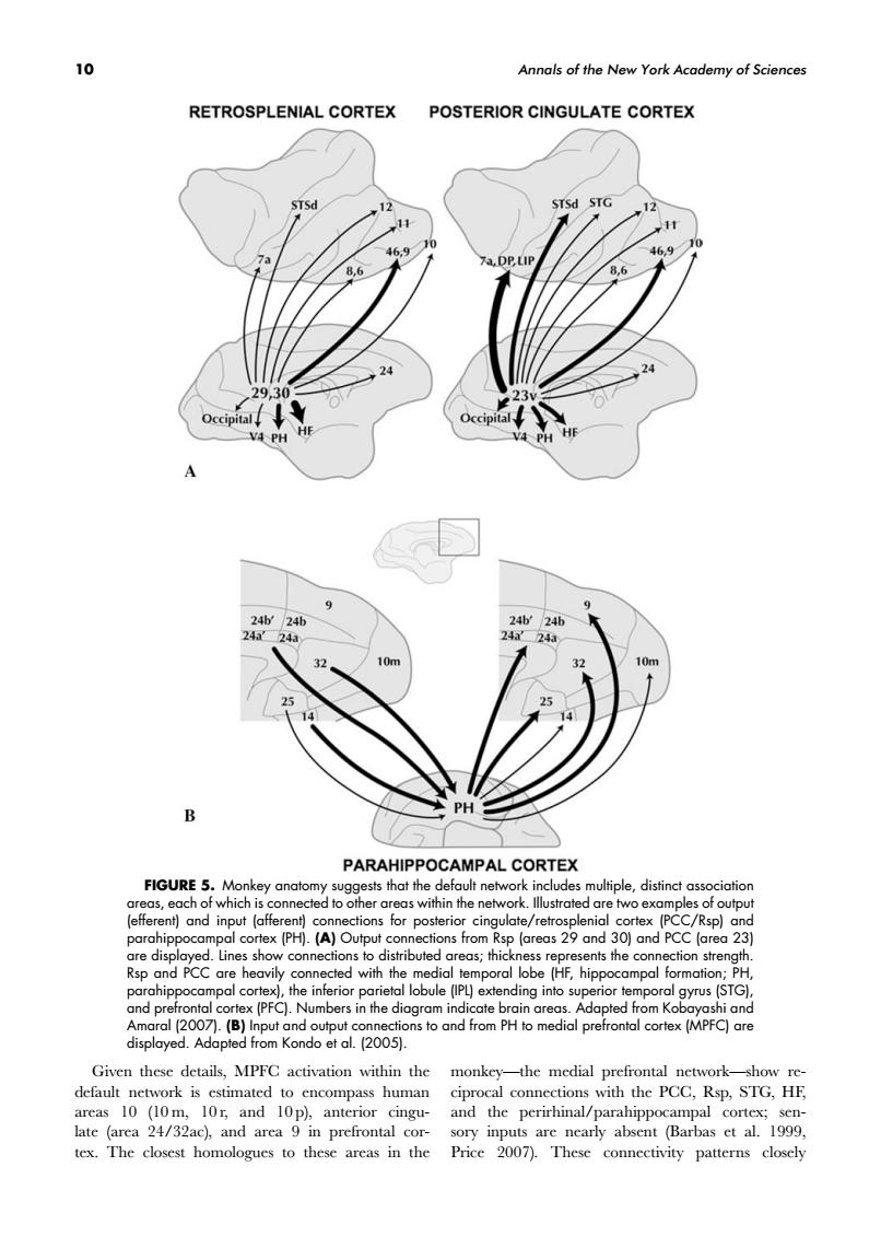
10 Annals of the New York Academy of Sciences RETROSPLENIAL CORTEX POSTERIOR CINGULATE CORTEX 24 PARAHIPPOCAMPAL CORTEX FIGURE 5.M ns t dis nts the co e.henecedwi obule (PL)exten Given these details,MPFC activation within the monkey-the medial prefrontal networkshow re default network is estimated to encompass human ciproc 100032ac to the areas in 10 Annals of the New York Academy of Sciences FIGURE 5. Monkey anatomy suggests that the default network includes multiple, distinct association areas, each of which is connected to other areas within the network. Illustrated are two examples of output (efferent) and input (afferent) connections for posterior cingulate/retrosplenial cortex (PCC/Rsp) and parahippocampal cortex (PH). (A) Output connections from Rsp (areas 29 and 30) and PCC (area 23) are displayed. Lines show connections to distributed areas; thickness represents the connection strength. Rsp and PCC are heavily connected with the medial temporal lobe (HF, hippocampal formation; PH, parahippocampal cortex), the inferior parietal lobule (IPL) extending into superior temporal gyrus (STG), and prefrontal cortex (PFC). Numbers in the diagram indicate brain areas. Adapted from Kobayashi and Amaral (2007). (B) Input and output connections to and from PH to medial prefrontal cortex (MPFC) are displayed. Adapted from Kondo et al. (2005). Given these details, MPFC activation within the default network is estimated to encompass human areas 10 (10 m, 10 r, and 10 p), anterior cingulate (area 24/32ac), and area 9 in prefrontal cortex. The closest homologues to these areas in the monkey—the medial prefrontal network—show reciprocal connections with the PCC, Rsp, STG, HF, and the perirhinal/parahippocampal cortex; sensory inputs are nearly absent (Barbas et al. 1999, Price 2007). These connectivity patterns closely