正在加载图片...

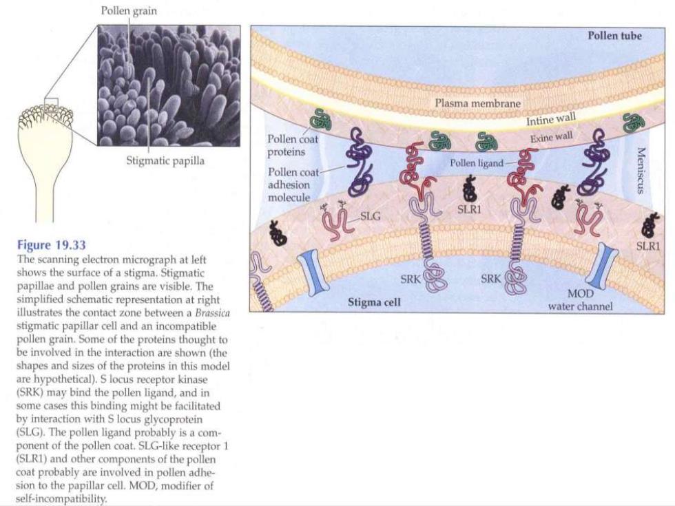
Pollen grain Pollen tube Plasma membrane Intine wall Pollen coat Exine wall Stigmatic papilla proteins Pollen ligand Pollen coat adhesion Meniscus molecule Figure 19.33 SLRI The scanning electron micrograph at left shows the surface of a stigma.Stigmatic papillae and pollen grains are visible.The SRK SRK simplified schematic representation at right MOD Stigma cell illustrates the contact zone between a Brassica water channel stigmatic papillar cell and an incompatible pollen grain.Some of the proteins thought to be involved in the interaction are shown(the shapes and sizes of the proteins in this model are hypothetical).S locus receptor kinase (SRK)may bind the pollen ligand,and in some cases this binding might be facilitated by interaction with S locus glycoprotein (SLG).The pollen ligand probably is a com ponent of the pollen coat.SLG-like receptor 1 (SLR1)and other components of the pollen coat probably are involved in pollen adhe- sion to the papillar cell.MOD,modifier of self-incompatibility