正在加载图片...

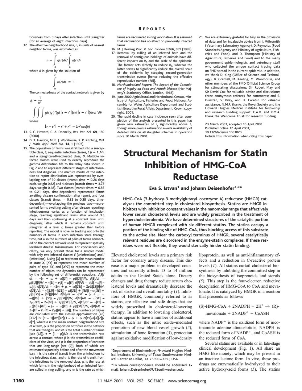
21.nd orm neighborhood size,n bor farn nR。and the eof the event x(r)dr =1 (0 rof the er Mal The of the contact network is given by 19. - Sa0ww9dr-rh-d-rhoa t2001 ng su and R. 20 ha the We when citing this pape Structural Mechanism for Statin Inhibition of HMG-CoA Reductase Eva S.Istvan'and Johann Deisenhofer12 thevesand are widelypres dues were no flexib they woud sterically hinder statin binding s in the Un State alone hesis of is ol levels and dra ally dec of HMGR to a that ar ed i (S)-HMG-CoA 2NADPH +2H*(R)- apy ion to low ter mevalonate+2NADP++CoASH where NADP is the oxidize of bone fomm inst oxidative moc -densit the reduced HM-i inactive lactor sep ring sulling and ss is the rate at which active hydroxy-acid forms (5).The statins 1160 11MAY2001 VOL 292 SCIENCE www.sciencemag.org tiousness from 3 days after infection until slaughter (for an average of eight infectious days). 12. The effective neighborhood size, n, in units of nearest neighbor farms, was estimated as n 5 E 0 ` g~r!dr/E 0 R g~r!dr where R is given by the solution of E 0 R k~r!dr 5 1 The connectedness of the contact network is given by f 5 1 n2 EEE g(r)g(r9)g(r 2 r9)/k(r 2 r9)drdr9du where r 2 r* 5 r 2 1 r9 2 2 2rr9cos(u) 13. S. C. Howard, C. A. Donnelly, Res. Vet. Sci. 69, 189 (2000). 14. D. T. Haydon, M. E. J. Woolhouse, R. P. Kitching, IMA J. Math. Appl. Med. Bio. 14, 1 (1997). 15. The population of farms was stratified into a susceptible class, S; sequential infection classes, I i (i 5 1..M); and a slaughtered/vaccinated class, D. Multiple infected classes were used to exactly reproduce the gamma distribution fits to the delay data shown in Fig. 2 and to represent different stages of infectiousness and diagnosis. The mixture model of the infection-to-report distribution was represented by overlapping sets of 30 classes (transit time 5 0.26 days each, weight 0.82) and 4 classes (transit times 5 3.73 days, weight 0.18). Two classes (transit times 5 0.85 to 0.21 days, time-dependent) represented farms awaiting disease confirmation after report, and four classes (transit times 5 0.82 to 0.38 days, timedependent)—overlapping the previous two—represented farms awaiting culling after disease reporting. Infectiousness varies as a function of incubation stage, reaching significant levels after around 3.5 days and then continuing at a constant level until diagnosis, after which it remains constant until slaughter at a level rI times greater than before reporting. The model is novel in tracking not only the numbers of farms in each infection state through time, but also the numbers of pairs of farms connected on the contact network used to represent spatially localized disease transmission. For conciseness and clarity, we only present those for a simpler model with only two infected classes: E (uninfectious) and I (infectious). Using [X] to represent the mean number in state X, [XY] to represent the mean number of pairs of type XY, and [XYZ] to represent the mean number of triples, the dynamics can be represented by the following set of differential equations: d[S]/ dt 5 –(t1m1v)[SI] – pb[S][I]/N, d[E]/dt 5 pb[S][I]/N 1 t[SI] – n[E] – m[EI], d[I]/dt 5 n[E] – s[I] – m[II], d[SS]/dt 5 –2(t1m1v)[SSI]–2pb[SS][I]/N, d[SE]/dt 5 t([SSI]–[ISE]) – m([SEI] 1 [ISE]) – v[ISE] 1 pb([SS]–[SE])[I]/N, d[SI]/dt 5 n[SE]–(t1m1 v)([ISI] 1 [SI]) – pb[SI][I]/N, d[EE]/dt 5 t[ISE] – 2m[EEI]–2n[EE] 1 2pb[SE][I]/N, d[EI]/dt 5 n[EE] – m([EI]1[IEI]) – (n1s)[EI] 1 pb[SI][I]/N, d[II]/dt 5 2n[EI]–2s[II]–2m([II] 1 [III]). The numbers of triples are calculated with the closure approximation (16) [XYZ] ' (n – 1)[XY][YZ](1 – f1f N[YY]/n[X][Z])/ n[Y], where n is the mean contact neighborhood size of a farm, f is the proportion of triples in the network that are triangles, and N is the total number of farms [see (12)]. t 5 (1 – p)b/n is the transmission rate across a contact, where b is the transmission coeffi- cient of the virus, and p is the proportion of contacts that are long-range [see (9)], both of which are estimated separately before and after the movement ban. n is the rate of transit from the uninfectious to the infectious class, and s is the rate of transit from the infectious to the removed class. m is the rate at which farms in the neighborhood of an infected farm are culled in ring culling, and v is the rate at which farms are vaccinated in ring vaccination. It is assumed that vaccination has no effect on previously infected farms. 16. M. J. Keeling, Proc. R. Soc. London B 266, 859 (1999). 17. Removal by culling of an infected herd and the removal of contiguous holdings of animals have different impacts on R0 and the scale of the epidemic. The former acts directly to reduce R0, whereas the latter serves to significantly reduce the overall scale of the epidemic by stopping second-generation transmission events [hence reducing the effective reproductive number (10)]. 18. Northumberland Report: The Report of the Committee of Inquiry on Food and Mouth Disease (Her Majesty’s Stationery Office, London, 1968). 19. June 2000 Agricultural and Horticultural Census, Ministry of Agriculture, Fisheries and Food, National Assembly for Wales Agriculture Department and Scottish Executive Rural Affairs Department; Crown copyright, 2001. 20. The rapid decline in case incidence seen after completion of the analysis presented in this paper has given new estimates of rI significantly above 1, though more precise estimation awaits availability of detailed data on all slaughter schemes in operation since 30 March 2001. 21. We are extremely grateful for help in the provision of data and for invaluable advice from J. Wilesmith (Veterinary Laboratory Agency), D. Reynolds (Food Standards Agency and Ministry of Agriculture, Fisheries and Food), and D. Thompson (Ministry of Agriculture, Fisheries and Food) and to the many government epidemiologists and veterinary staff who collected the unique contact tracing data on FMD spread in the current epidemic. In addition, we thank D. King (Office of Science and Technology), B. Grenfell, M. Keeling, M. Woolhouse, and other members of the FMD Official Science Group for stimulating discussions; Sir Robert May and Sir David Cox for valuable advice and discussions; three anonymous referees for comments; and S. Dunstan, S. Riley, and H. Carabin for valuable assistance. N.M.F. thanks the Royal Society and the Howard Hughes Medical Institute for fellowship and research funding support. C.A.D. and R.M.A. thank the Wellcome Trust for research funding. 23 March 2001; accepted 10 April 2001 Published online 12 April 2001; 10.1126/science.1061020 Include this information when citing this paper. Structural Mechanism for Statin Inhibition of HMG-CoA Reductase Eva S. Istvan1 and Johann Deisenhofer1,2* HMG-CoA (3-hydroxy-3-methylglutaryl–coenzyme A) reductase (HMGR) catalyzes the committed step in cholesterol biosynthesis. Statins are HMGR inhibitors with inhibition constant values in the nanomolar range that effectively lower serum cholesterol levels and are widely prescribed in the treatment of hypercholesterolemia. We have determined structures of the catalytic portion of human HMGR complexed with six different statins. The statins occupy a portion of the binding site of HMG-CoA, thus blocking access of this substrate to the active site. Near the carboxyl terminus of HMGR, several catalytically relevant residues are disordered in the enzyme-statin complexes. If these residues were not flexible, they would sterically hinder statin binding. Elevated cholesterol levels are a primary risk factor for coronary artery disease. This disease is a major problem in developed countries and currently affects 13 to 14 million adults in the United States alone. Dietary changes and drug therapy reduce serum cholesterol levels and dramatically decrease the risk of stroke and overall mortality (1). Inhibitors of HMGR, commonly referred to as statins, are effective and safe drugs that are widely prescribed in cholesterol-lowering therapy. In addition to lowering cholesterol, statins appear to have a number of additional effects, such as the nitric oxide–mediated promotion of new blood vessel growth (2), stimulation of bone formation (3), protection against oxidative modification of low-density lipoprotein, as well as anti-inflammatory effects and a reduction in C-reactive protein levels (4). All statins curtail cholesterol biosynthesis by inhibiting the committed step in the biosynthesis of isoprenoids and sterols (5). This step is the four-electron reductive deacylation of HMG-CoA to CoA and mevalonate. It is catalyzed by HMGR in a reaction that proceeds as follows (S)-HMG-CoA 1 2NADPH 1 2H1 3 (R)- mevalonate 1 2NADP1 1 CoASH where NADP1 is the oxidized form of nicotinamide adenine dinucelotide, NADPH is the reduced form of NADP1, and CoASH is the reduced form of CoA. Several statins are available or in late-stage clinical development (Fig. 1). All share an HMG-like moiety, which may be present in an inactive lactone form. In vivo, these prodrugs are enzymatically hydrolyzed to their active hydroxy-acid forms (5). The statins 1 Department of Biochemistry, 2 Howard Hughes Medical Institute, University of Texas Southwestern Medical Center at Dallas, TX 75390–9050, USA. *To whom correspondence should be addressed. Email: Johann.Deisenhofer@UTSouthwestern.edu R EPORTS 1160 11 MAY 2001 VOL 292 SCIENCE www.sciencemag.org