正在加载图片...
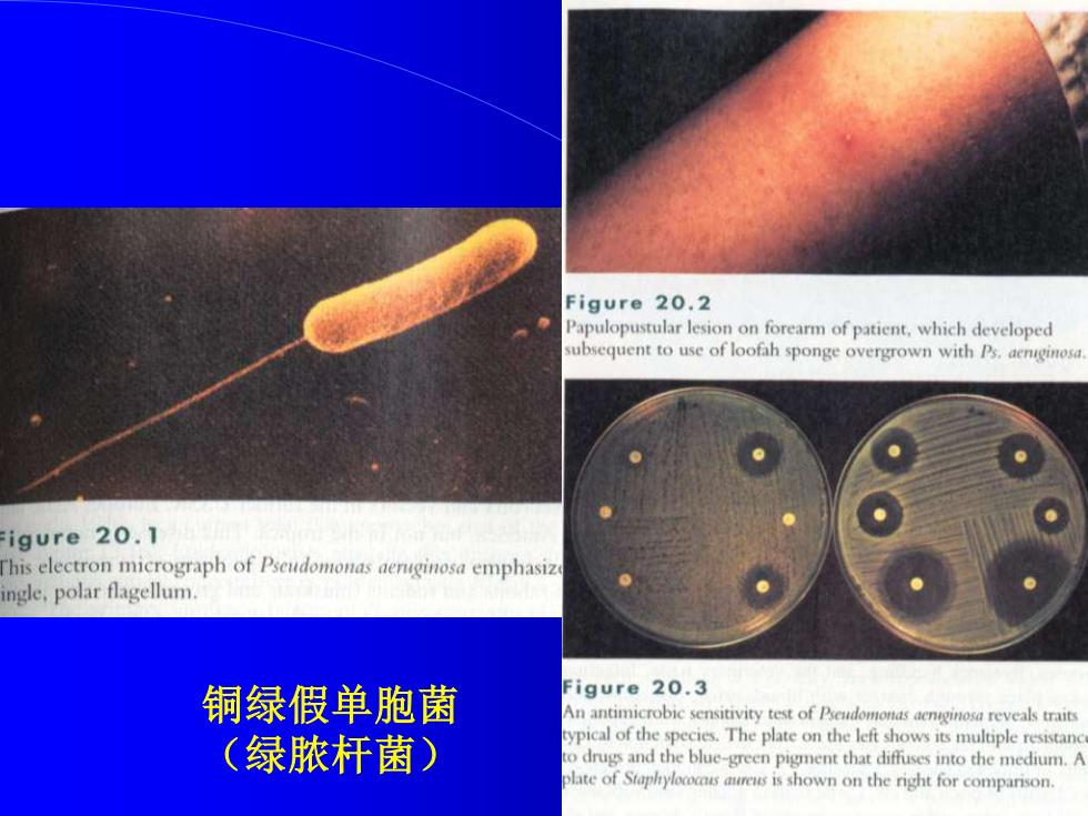
Figure 20.2 Papulopustular lesion on forearm of patient,which developed subsequent to use of loofah sponge overgrown with Ps.aenginos. igure 20.1 his electron micrograph of Pseudomonas aenginosa emphasiz ngle,polar flagellum. 铜绿假单胞菌 Figure 20.3 An antimicrobic sensitivity test of Pseudomonas aenginosa reveals traits (绿脓杆菌) ypical of the species.The plate on the left shows its multiple resistanc o drugs and the blue-green pigment that diffuses into the medium.A late of Staplryloccsres is shown on the right for comparison.铜绿假单胞菌 (绿脓杆菌)