正在加载图片...
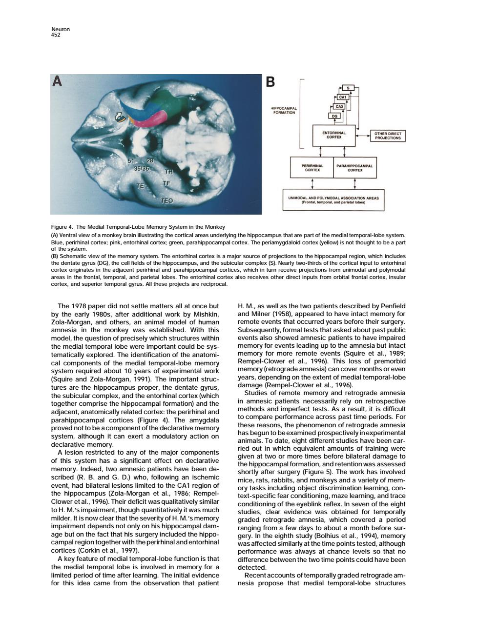
B The Medial Te ral-Lobe Me System in the Monke B)Schemat urce of p x also receives other direc inputs from orbital frontal cortex,insul The 1978p paper did not settle matters all at once but H.M..as well as the two patients described by Penfield work by te events that occum rs h re their sur del.th 8nieqgegy,ormale tha al which s red he medial temporal important ng up t et al ut inge al components of the nedial temporal-lobe me y(retro sial ca Squire and zola-Morg al temporal-lobe the hippoca npus proper.the d tate memory and ograde amne ethe mpal formati erfect tests past is dif the phenomenon of retr ystem,although it can exert a modulatory action on rade amnes nt st th major nponents at t re bilat ge t this heer n de ry(Figure 5).The cribe (R.B.an to the Zola-M gan t al. t d 1986 o H.M.'s impairment,though quantitativ it was gshnepenashol on his h ghth 994 cs Corkin et al. function is tha ce was at cha ce tha ume po the medial temporal lo e is involy in for a have becn for this idea came from the vation that patient Neuron 452 Figure 4. The Medial Temporal-Lobe Memory System in the Monkey (A) Ventral view of a monkey brain illustrating the cortical areas underlying the hippocampus that are part of the medial temporal-lobe system. Blue, perirhinal cortex; pink, entorhinal cortex; green, parahippocampal cortex. The periamygdaloid cortex (yellow) is not thought to be a part of the system. (B) Schematic view of the memory system. The entorhinal cortex is a major source of projections to the hippocampal region, which includes the dentate gyrus (DG), the cell fields of the hippocampus, and the subicular complex (S). Nearly two-thirds of the cortical input to entorhinal cortex originates in the adjacent perirhinal and parahippocampal cortices, which in turn receive projections from unimodal and polymodal areas in the frontal, temporal, and parietal lobes. The entorhinal cortex also receives other direct inputs from orbital frontal cortex, insular cortex, and superior temporal gyrus. All these projects are reciprocal. The 1978 paper did not settle matters all at once but H. M., as well as the two patients described by Penfield by the early 1980s, after additional work by Mishkin, and Milner (1958), appeared to have intact memory for Zola-Morgan, and others, an animal model of human remote events that occurred years before their surgery. amnesia in the monkey was established. With this Subsequently, formal tests that asked about past public model, the question of precisely which structures within events also showed amnesic patients to have impaired memory for events leading up to the amnesia but intact the medial temporal lobe were important could be sysmemory for more remote events (Squire et al., 1989; tematically explored. The identification of the anatomical components of the medial temporal-lobe memory Rempel-Clower et al., 1996). This loss of premorbid memory (retrograde amnesia) can cover months or even system required about 10 years of experimental work years, depending on the extent of medial temporal-lobe (Squire and Zola-Morgan, 1991). The important struc- damage (Rempel-Clower et al., 1996). tures are the hippocampus proper, the dentate gyrus, Studies of remote memory and retrograde amnesia the subicular complex, and the entorhinal cortex (which in amnesic patients necessarily rely on retrospective together comprise the hippocampal formation) and the methods and imperfect tests. As a result, it is difficult adjacent, anatomically related cortex: the perirhinal and to compare performance across past time periods. For parahippocampal cortices (Figure 4). The amygdala these reasons, the phenomenon of retrograde amnesia proved not to be a component of the declarative memory has begun to beexamined prospectively inexperimental system, although it can exert a modulatory action on animals. To date, eight different studies have been car- declarative memory. ried out in which equivalent amounts of training were A lesion restricted to any of the major components given at two or more times before bilateral damage to of this system has a significant effect on declarative the hippocampal formation, and retention was assessed memory. Indeed, two amnesic patients have been de- shortly after surgery (Figure 5). The work has involved scribed (R. B. and G. D.) who, following an ischemic mice, rats, rabbits, and monkeys and a variety of mem- event, had bilateral lesions limited to the CA1 region of ory tasks including object discrimination learning, con- the hippocampus (Zola-Morgan et al., 1986; Rempel- text-specific fear conditioning, maze learning, and trace Clower et al., 1996). Their deficit was qualitatively similar conditioning of the eyeblink reflex. In seven of the eight to H. M.’s impairment, though quantitatively it was much studies, clear evidence was obtained for temporally milder. It is now clear that the severity of H. M.’s memory graded retrograde amnesia, which covered a period impairment depends not only on his hippocampal dam- ranging from a few days to about a month before surage but on the fact that his surgery included the hippo- gery. In the eighth study (Bolhius et al., 1994), memory campal region togetherwith theperirhinal and entorhinal was affected similarly at the time points tested, although cortices (Corkin et al., 1997). performance was always at chance levels so that no A key feature of medial temporal-lobe function is that difference between the two time points could have been the medial temporal lobe is involved in memory for a detected. limited period of time after learning. The initial evidence Recent accounts of temporally graded retrograde amfor this idea came from the observation that patient nesia propose that medial temporal-lobe structures