正在加载图片...
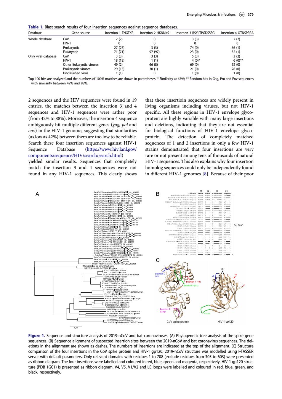
Emerging Microbes&nfections379 Table 1.Blast search resus of four atabas Eukaryotic viruses 13 dtot10%maeseownnp时emtei'snbi内yt6xRnnhsnogon2qens ces and the HIV sec e found in 19 that these insertioin entries the matches living sequences and HIV-1 sequences were rather poor specific.All these regions in HIV-1 envelope glyco (from 42%to 88%).Moreover,the insertion 4 sequence t genes (ag pol and ng tha Hivey are of Search these four insertion sequences against HIV-1 sequences of 1 and 2 insertions in only a few HIV-1 Sequence Database (https://www.hiv.lanL.gov/ strains demonstrated that four insertions are very nents/ r res andnces that present mong tens of thousands of natura sequences.Thi found in any HIV-1 seg ences.This clearly shows in different HIV-I genomes 8).Because of their poo CoV spike prot HN-1 gp120 Figure 1.Sequence and str uctureaysisof19-Con bat.(A)of the etions in the anment are sho ated at the top of the alig nt (o)5 Co e pr ng I-TASSE were lab black respectively. 2 sequences and the HIV sequences were found in 19 entries, the matches between the insertion 3 and 4 sequences and HIV-1 sequences were rather poor (from 42% to 88%). Moreover, the insertion 4 sequence ambiguously hit multiple different genes (gag, pol and env) in the HIV-1 genome, suggesting that similarities (as low as 42%) between them are too low to be reliable. Search these four insertion sequences against HIV-1 Sequence Database (https://www.hiv.lanl.gov/ components/sequence/HIV/search/search.html) yielded similar results. Sequences that completely match the insertion 3 and 4 sequences were not found in any HIV-1 sequences. This clearly shows that these insertioin sequences are widely present in living organisms including viruses, but not HIV-1 specific. All these regions in HIV-1 envelope glycoprotein are highly variable with many large insertions and deletions, indicating that they are not essential for biological functions of HIV-1 envelope glycoprotein. The detection of completely matched sequences of 1 and 2 insertions in only a few HIV-1 strains demonstrated that four insertions are very rare or not present among tens of thousands of natural HIV-1 sequences. This also explains why four insertion homolog sequences could only be independently found in different HIV-1 genomes [8]. Because of their poor Table 1. Blast search results of four insertion sequences against sequence databases. Database Gene source Insertion 1 TNGTKR Insertion 2 HKNNKS Insertion 3 RSYLTPGDSSSG Insertion 4 QTNSPRRA Whole database CoV 2 (2) 0 3 (3) 2 (2) HIV-1 0 0 0 0 Prokaryotic 27 (27) 3 (3) 74 (0) 66 (1) Eukaryotic 71 (71) 97 (97) 23 (0) 32 (1) Only viral database CoV 3 (3) 3 (3) 5 (3) 3 (2) HIV-1 18 (18) 1 (1) 4 (0)* 6 (0)** Other Eukaryotic viruses 49 (2) 66 (8) 69 (0) 62 (0) Prokaryotic viruses 29 (13) 30 (1) 21 (0) 28 (0) Unclassified virus 1 (1) 0 1 (0) 1 (0) Top 100 hits are analyzed and the numbers of 100% matches are shown in parentheses. * Similarity at 67%; ** Random hits in Gag, Pro and Env sequences with similarity between 42% and 88%. Figure 1. Sequence and structure analysis of 2019-nCoV and bat coronaviruses. (A) Phylogenetic tree analysis of the spike gene sequences. (B) Sequence alignment of suspected insertion sites between the 2019-nCoV and bat coronavirus sequences. The deletions in the alignment are shown as dashes. The numbers of insertions are indicated at the top of the alignment. (C) Structure comparison of the four insertions in the CoV spike protein and HIV-1 gp120. 2019-nCoV structure was modelled using I-TASSER server with default parameters. Only relevant domains with residues 1 to 708 (exclude residues from 305 to 603) were presented as ribbon diagram. The four insertions were labelled and coloured in red, blue, green and magenta, respectively. HIV-1 gp120 structure (PDB 1GC1) is presented as ribbon diagram. V4, V5, V1/V2 and LE loops were labelled and coloured in red, blue, green, and black, respectively. Emerging Microbes & Infections 379