正在加载图片...
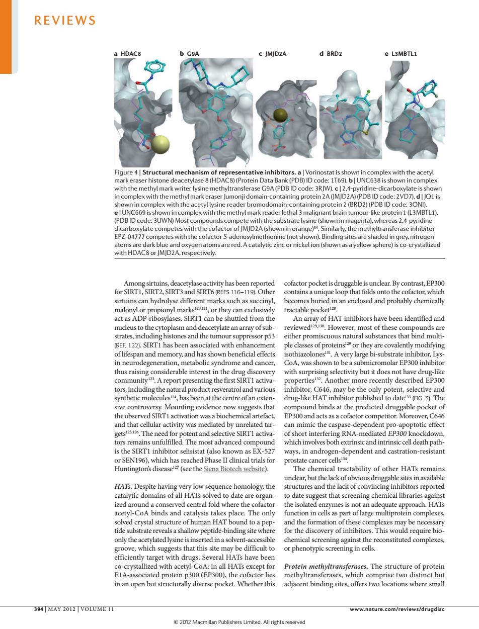
REVIEWS a HDAC c JMJD2A d BRD2 inhibitors.a|Vorir with the acety R ng P with the (BRD2)(PDBID BON BIDcode:3UWN)Most ke prot P7-047cmwith the ey nitr ih HDAC&IMIDZA s are red.A catalytic zinc or nick ion(st -cryst cofactor pocket is druggable is inclear.By contrast,EP30 sirtuins can hvdrolyse different marks such as su tractable pocke HAT inhibitors hav lasm and deacetvlate an arravofsub evie However,most of these nds ar substances hat bind muti and memory and has shov A ve metabolic syn ome and cancer, was shown P300 ommunity Areport presenting the first S ently described EP300 hndtdoShcaurlprodtctrcevecrtrolandarniou may be th sts that edicted druggable pocket of n was a t O a cts as a cofa r competitor. th The nce ent and sele RNA- ckdo pro ate cance e Siena Biotech website) of othe HATs rem ence l omology,the tructures and the lack of convincing ink s reporte da are organ st that scre ning c lated ety-CoA binds and alysis ta es place The only fun as par tof large complexe tide-binding quire bio only the ed in a sol hicalscreeningagainstth reconstituted complexes or phenotypic screening in with acetyl- Protein structur of proteir in an open but structurally dive pocket.Whether this adiacent binding sites.offers twp locations where small 394 MAY 2012 VOLUME 11 www.nature.com/reviews/drugdisc 2012 Macmillan Publishers Limited All riehts reservee Nature Reviews | Drug Discovery a HDAC8 b G9A c JMJD2A d BRD2 e L3MBTL1 Among sirtuins, deacetylase activity has been reported for SIRT1, SIRT2, SIRT3 and SIRT6 (REFS 116–119). Other sirtuins can hydrolyse different marks such as succinyl, malonyl or propionyl marks120,121, or they can exclusively act as ADP-ribosylases. SIRT1 can be shuttled from the nucleus to the cytoplasm and deacetylate an array of substrates, including histones and the tumour suppressor p53 (REF. 122). SIRT1 has been associated with enhancement of lifespan and memory, and has shown beneficial effects in neurodegeneration, metabolic syndrome and cancer, thus raising considerable interest in the drug discovery community123. A report presenting the first SIRT1 activators, including the natural product resveratrol and various synthetic molecules124, has been at the centre of an extensive controversy. Mounting evidence now suggests that the observed SIRT1 activation was a biochemical artefact, and that cellular activity was mediated by unrelated targets125,126. The need for potent and selective SIRT1 activators remains unfulfilled. The most advanced compound is the SIRT1 inhibitor selisistat (also known as EX-527 or SEN196), which has reached Phase II clinical trials for Huntington’s disease127 (see the Siena Biotech website). HATs. Despite having very low sequence homology, the catalytic domains of all HATs solved to date are organized around a conserved central fold where the cofactor acetyl-CoA binds and catalysis takes place. The only solved crystal structure of human HAT bound to a peptide substrate reveals a shallow peptide-binding site where only the acetylated lysine is inserted in a solvent-accessible groove, which suggests that this site may be difficult to efficiently target with drugs. Several HATs have been co-crystallized with acetyl-CoA: in all HATs except for E1A-associated protein p300 (EP300), the cofactor lies in an open but structurally diverse pocket. Whether this cofactor pocket is druggable is unclear. By contrast, EP300 contains a unique loop that folds onto the cofactor, which becomes buried in an enclosed and probably chemically tractable pocket128. An array of HAT inhibitors have been identified and reviewed129,130. However, most of these compounds are either promiscuous natural substances that bind multiple classes of proteins129 or they are covalently modifying isothiazolones131. A very large bi-substrate inhibitor, LysCoA, was shown to be a submicromolar EP300 inhibitor with surprising selectivity but it does not have drug-like properties132. Another more recently described EP300 inhibitor, C646, may be the only potent, selective and drug-like HAT inhibitor published to date133 (FIG. 3). The compound binds at the predicted druggable pocket of EP300 and acts as a cofactor competitor. Moreover, C646 can mimic the caspase-dependent pro-apoptotic effect of short interfering RNA-mediated EP300 knockdown, which involves both extrinsic and intrinsic cell death pathways, in androgen-dependent and castration-resistant prostate cancer cells134. The chemical tractability of other HATs remains unclear, but the lack of obvious druggable sites in available structures and the lack of convincing inhibitors reported to date suggest that screening chemical libraries against the isolated enzymes is not an adequate approach. HATs function in cells as part of large multiprotein complexes, and the formation of these complexes may be necessary for the discovery of inhibitors. This would require biochemical screening against the reconstituted complexes, or phenotypic screening in cells. Protein methyltransferases. The structure of protein methyltransferases, which comprise two distinct but adjacent binding sites, offers two locations where small Figure 4 | Structural mechanism of representative inhibitors. a | Vorinostat is shown in complex with the acetyl mark eraser histone deacetylase 8 (HDAC8) (Protein Data Bank (PDB) ID code: 1T69). b | UNC638 is shown in complex with the methyl mark writer lysine methyltransferase G9A (PDB ID code: 3RJW). c | 2,4‑pyridine-dicarboxylate is shown in complex with the methyl mark eraser Jumonji domain-containing protein 2A (JMJD2A) (PDB ID code: 2VD7). d | JQ1 is shown in complex with the acetyl lysine reader bromodomain-containing protein 2 (BRD2) (PDB ID code: 3ONI). e | UNC669 is shown in complex with the methyl mark reader lethal 3 malignant brain tumour-like protein 1 (L3MBTL1). (PDB ID code: 3UWN) Most compounds compete with the substrate lysine (shown in magenta), whereas 2,4-pyridinedicarboxylate competes with the cofactor of JMJD2A (shown in orange)34. Similarly, the methyltransferase inhibitor EPZ-04777 competes with the cofactor S-adenosylmethionine (not shown). Binding sites are shaded in grey, nitrogen atoms are dark blue and oxygen atoms are red. A catalytic zinc or nickel ion (shown as a yellow sphere) is co-crystallized with HDAC8 or JMJD2A, respectively. REVIEWS 394 | MAY 2012 | VOLUME 11 www.nature.com/reviews/drugdisc © 2012 Macmillan Publishers Limited. All rights reserved