正在加载图片...

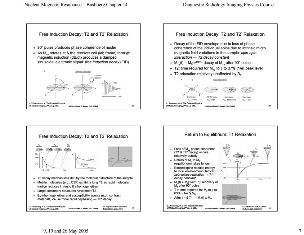
Nuclear Magnetic Resonance-Bushberg Chapter 14 Diagnostic Radiology Imaging Physics Course Free Induction Decay:T2 and T2'Relaxation Free Induction Decay:T2 and T2'Relaxation Decay of the FID envelope due to loss of phase ◆9o°oulse produces phase coherence of nuclei coherence of the individual spins due to As Mrotates at fo the receiver coil (lab frame)through magnetic field variations in the sample:spin-spin magnetic induction(dB/dt)produces a damped interaction-T2 decay constant sinusoidal electronic signal:free induction decay(FID) ÷M)-M,ewr2a:decay of M after90°pulse T2:time required for Mto to 37%(1/e)peak level o9m T2 relaxation relatively unaffected by Bo Bontno fram 咖 色W Free Induction Decay:T2 and T2'Relaxation Return to Equilibrium:T1 Relaxation cay)occurs elatively quickly spin-lattice relaxationT1 .T2 decay mechanisms det.by the molecular structure of the sample .Mobile molecules (e.g.CSF)exhibit a long T2 as rapid molecular motion reduces intrinsic B inhomogeneities Large stationary structures have short t2 ◆Aftert=5T1一M)=Mo 品ma 9.19and26May2005 >Nuclear Magnetic Resonance – Bushberg Chapter 14 Diagnostic Radiology Imaging Physics Course 9, 19 and 26 May 2005 7 © UW and Brent K. Stewart, PhD, DABMP 25 Free Induction Decay: T2 and T2* Relaxation 90° pulse produces phase coherence of nuclei pulse produces phase coherence of nuclei As Mxy rotates at f rotates at f0 the receiver coil (lab frame) through the receiver coil (lab frame) through magnetic induction (dB/dt) produces a damped magnetic induction (dB/dt) produces a damped sinusoidal electronic signal: sinusoidal electronic signal: free induction decay (FID) c.f. Bushberg, et al. The Essential Physics of Medical Imaging, 2nd ed., p. 385. © UW and Brent K. Stewart, PhD, DABMP 26 Free Induction Decay: T2 and T2* Relaxation Decay of the FID envelope due to loss of phase Decay of the FID envelope due to loss of phase coherence of the individual spins due to intrinsic micro coherence of the individual spins due to intrinsic micro magnetic field variations in the sample: magnetic field variations in the sample: spin-spin interaction interaction → T2 decay constant T2 decay constant Mxy(t) = M0e-(t/T2): decay of Mxy after 90° pulse T2: time required for Mxy to ↓ to 37% (1/e) peak level to 37% (1/e) peak level T2 relaxation relatively unaffected by B0 c.f. Bushberg, et al. The Essential Physics of Medical Imaging, 2nd ed., p. 385. © UW and Brent K. Stewart, PhD, DABMP 27 Free Induction Decay: T2 and T2* Relaxation T2 decay mechanisms det. by the molecular structure of the sampl T2 decay mechanisms det. by the molecular structure of the sample Mobile molecules (e.g., CSF) exhibit a long T2 as rapid molecular motion reduces intrinsic B inhomogeneities Large, stationary structures have short T2 B0 inhomogeneities and susceptibility agents (e.g., contrast inhomogeneities and susceptibility agents (e.g., contrast materials) cause more rapid dephasing materials) cause more rapid dephasing → T2* decay c.f. Bushberg, et al. The Essential Physics of Medical Imaging, 2nd ed., p. 386. c.f. http://www.hull.ac.uk/mri /lectures/gpl_page.html © UW and Brent K. Stewart, PhD, DABMP 28 Return to Equilibr Return to Equilibrium: T1 Relaxation Loss of Mxy phase coherence phase coherence (T2 & T2* decay) occurs (T2 & T2* decay) occurs relatively quickly Return of Mz to M0 (equilibrium) takes longer (equilibrium) takes longer Excited spins release energy Excited spins release energy to local environment (‘lattice’): to local environment (‘lattice’): spin-lattice relaxation lattice relaxation → T1 decay constant decay constant Mz(t) = M0[1-e-(t/T1)]: recovery of : recovery of Mz after 90° pulse T1: time required for Mz to ↑ to 63%: (1-e-1) M0 After t = 5 T1 After t = 5 T1 → Mz(t) ≅ M0 c.f. Bushberg, et al. The Essential Physics of Medical Imaging, 2nd ed., p. 387. c.f. http://www.hull.ac.uk/mri /lectures/gpl_page.html