正在加载图片...
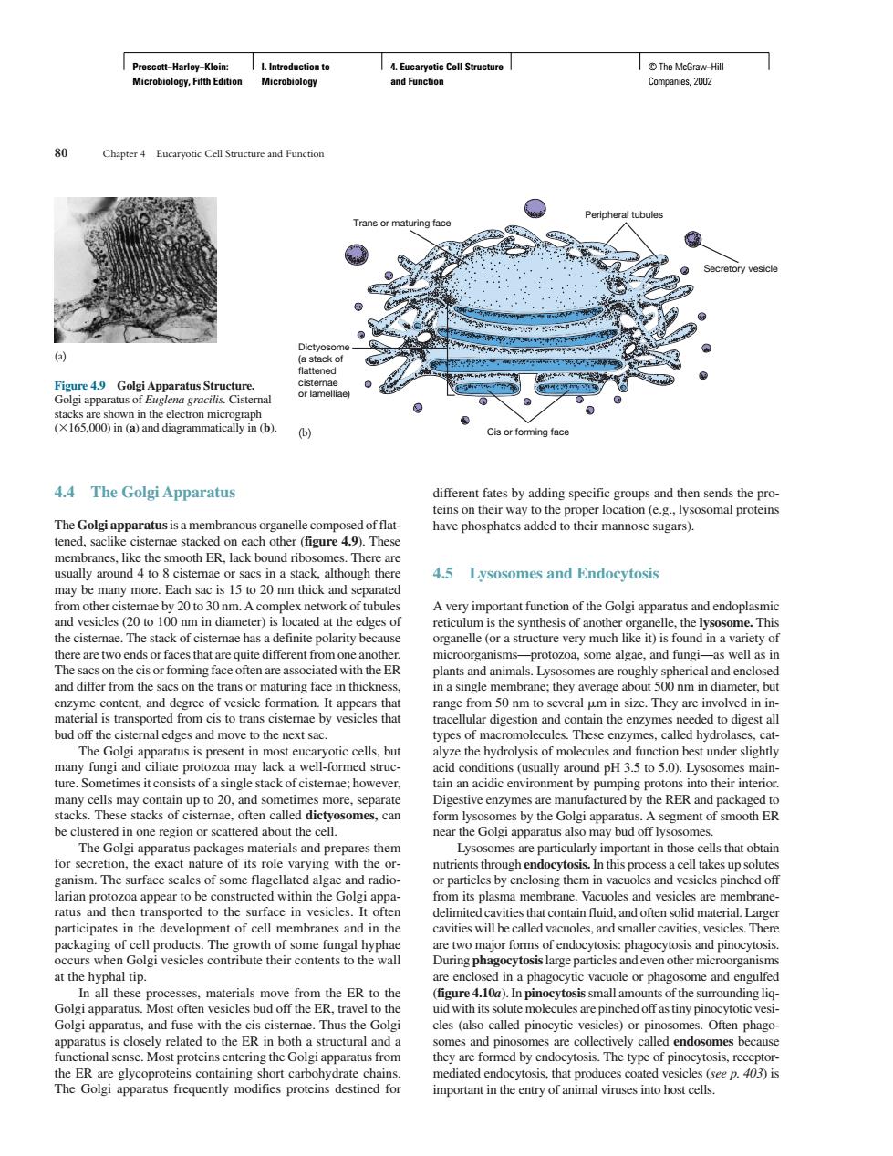
(165,000)in (and diagrammatically n(b) 4.4 The Golgi Apparatus different fates by adding specific and then sends the eins on their way to thep nave phosphates added to their mannose sugars). smooth ER.lack bound rib The may be many more.Each sac is 15 to 20 nm thick and s 4.5 Lysosomes and Endocytosis Avery important function of the Golgi apparatus and endoplasmic the cistemae.The stack of cisternae has a definite polarity be some algae and n a single membrane:they average ab out 500nm in diameter.but size.They are involved in i etimes it consis sts of a single stack of cistemae:however tain an acidic environment by pumping protons s into their interior ER The surtace with the s through endoc ossIn this process a cell takes up solute les of some rlagell ac and radic y enclosing them in vacuo esicles pinc and then ce in ities that contain fuid,and mat al.Lan participate in th ler cavitie 5m a the er to the 410 Most often ves cles bud off the ER.travel to the th its s ytotic ves from ed by ePrescott−Harley−Klein: Microbiology, Fifth Edition I. Introduction to Microbiology 4. Eucaryotic Cell Structure and Function © The McGraw−Hill Companies, 2002 4.4 The Golgi Apparatus The Golgi apparatusis a membranous organelle composed of flattened, saclike cisternae stacked on each other (figure 4.9). These membranes, like the smooth ER, lack bound ribosomes. There are usually around 4 to 8 cisternae or sacs in a stack, although there may be many more. Each sac is 15 to 20 nm thick and separated from other cisternae by 20 to 30 nm. A complex network of tubules and vesicles (20 to 100 nm in diameter) is located at the edges of the cisternae. The stack of cisternae has a definite polarity because there are two ends or faces that are quite different from one another. The sacs on the cis or forming face often are associated with the ER and differ from the sacs on the trans or maturing face in thickness, enzyme content, and degree of vesicle formation. It appears that material is transported from cis to trans cisternae by vesicles that bud off the cisternal edges and move to the next sac. The Golgi apparatus is present in most eucaryotic cells, but many fungi and ciliate protozoa may lack a well-formed structure. Sometimes it consists of a single stack of cisternae; however, many cells may contain up to 20, and sometimes more, separate stacks. These stacks of cisternae, often called dictyosomes, can be clustered in one region or scattered about the cell. The Golgi apparatus packages materials and prepares them for secretion, the exact nature of its role varying with the organism. The surface scales of some flagellated algae and radiolarian protozoa appear to be constructed within the Golgi apparatus and then transported to the surface in vesicles. It often participates in the development of cell membranes and in the packaging of cell products. The growth of some fungal hyphae occurs when Golgi vesicles contribute their contents to the wall at the hyphal tip. In all these processes, materials move from the ER to the Golgi apparatus. Most often vesicles bud off the ER, travel to the Golgi apparatus, and fuse with the cis cisternae. Thus the Golgi apparatus is closely related to the ER in both a structural and a functional sense. Most proteins entering the Golgi apparatus from the ER are glycoproteins containing short carbohydrate chains. The Golgi apparatus frequently modifies proteins destined for different fates by adding specific groups and then sends the proteins on their way to the proper location (e.g., lysosomal proteins have phosphates added to their mannose sugars). 4.5 Lysosomes and Endocytosis A very important function of the Golgi apparatus and endoplasmic reticulum is the synthesis of another organelle, the lysosome. This organelle (or a structure very much like it) is found in a variety of microorganisms—protozoa, some algae, and fungi—as well as in plants and animals. Lysosomes are roughly spherical and enclosed in a single membrane; they average about 500 nm in diameter, but range from 50 nm to several m in size. They are involved in intracellular digestion and contain the enzymes needed to digest all types of macromolecules. These enzymes, called hydrolases, catalyze the hydrolysis of molecules and function best under slightly acid conditions (usually around pH 3.5 to 5.0). Lysosomes maintain an acidic environment by pumping protons into their interior. Digestive enzymes are manufactured by the RER and packaged to form lysosomes by the Golgi apparatus. A segment of smooth ER near the Golgi apparatus also may bud off lysosomes. Lysosomes are particularly important in those cells that obtain nutrients through endocytosis. In this process a cell takes up solutes or particles by enclosing them in vacuoles and vesicles pinched off from its plasma membrane. Vacuoles and vesicles are membranedelimited cavities that contain fluid, and often solid material. Larger cavities will be called vacuoles, and smaller cavities, vesicles. There are two major forms of endocytosis: phagocytosis and pinocytosis. During phagocytosislarge particles and even other microorganisms are enclosed in a phagocytic vacuole or phagosome and engulfed (figure 4.10a). In pinocytosissmall amounts of the surrounding liquid with its solute molecules are pinched off as tiny pinocytotic vesicles (also called pinocytic vesicles) or pinosomes. Often phagosomes and pinosomes are collectively called endosomes because they are formed by endocytosis. The type of pinocytosis, receptormediated endocytosis, that produces coated vesicles (see p. 403) is important in the entry of animal viruses into host cells. 80 Chapter 4 Eucaryotic Cell Structure and Function Dictyosome (a stack of flattened cisternae or lamelliae) Trans or maturing face Peripheral tubules Secretory vesicle Cis or forming face Figure 4.9 Golgi Apparatus Structure. Golgi apparatus of Euglena gracilis. Cisternal stacks are shown in the electron micrograph (165,000) in (a) and diagrammatically in (b). (a) (b)�