正在加载图片...

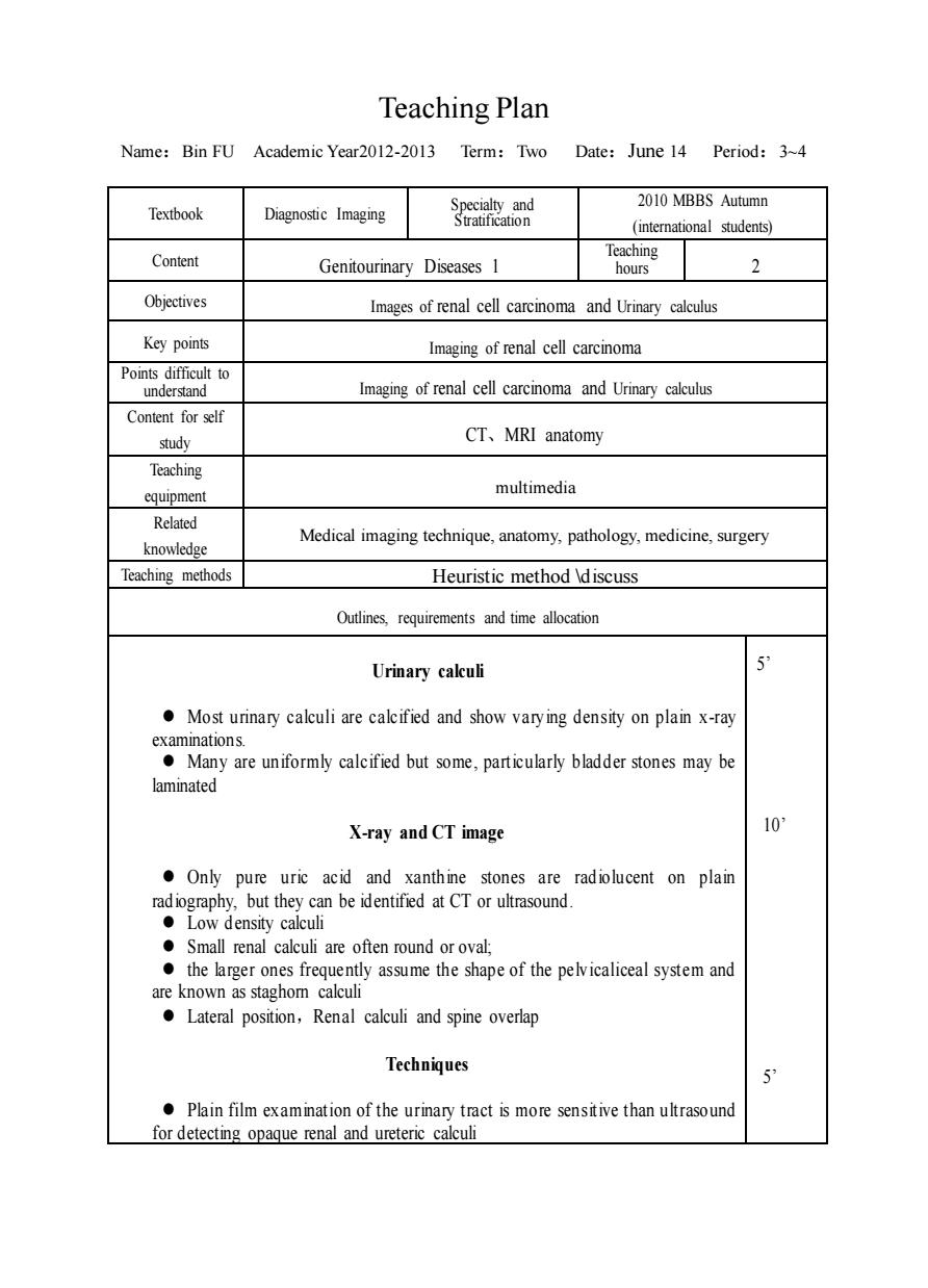
Teaching Plan Name:Bin FU Academic Year2012-2013 Term:Two Date:June 14 Period:3-4 2010 MBBS Autumn Textbook Diagnostic Imaging ion Content Genitourinary Diseases 1 Teaching 2 Objectives Images of renal cell carcinoma and Urinary calculus Key points Imaging of renal cell carcinoma Points difficult to under Imaging of renal cell carcinoma and Urinary calculus Content for self study CT、MRI anatomy Teaching equipment multimedia Related knowledge Medical imaging technique,anatomy,pathology,medicine,surgery Teaching methods Heuristic method discuss Outlines,requirements and time allocation Urinary cakuli Most urinary calculi are calcified and show vary ing density on plain x-ray e y are uniformly calcified but some.particularly bladder stones may be laminated X-ray and CT image 10 Only pure uric acid and xanthine stones are radiolucent on plain radiography,but they can be identified at CT or ultrasound. ●Low density calculi ●Small rena calculi are often round oroval ●the larger on are known as staghom Lateral position,Renal calculi and spine overlap Techniques Plain film examination of the urinary tract is more sensitive than ultrasound for detecting opaque renal and uretene calcul Teaching Plan Name:Bin FU Academic Year2012-2013 Term:Two Date:June 14 Period:3~4 Textbook Diagnostic Imaging Specialty and Stratification 2010 MBBS Autumn (international students) Content Genitourinary Diseases 1 Teaching hours 2 Objectives Images of renal cell carcinoma and Urinary calculus Key points Imaging of renal cell carcinoma Points difficult to understand Imaging of renal cell carcinoma and Urinary calculus Content for self study CT、MRI anatomy Teaching equipment multimedia Related knowledge Medical imaging technique, anatomy, pathology, medicine, surgery Teaching methods Heuristic method \discuss Outlines, requirements and time allocation Urinary calculi ⚫ Most urinary calculi are calcified and show varying density on plain x-ray examinations. ⚫ Many are uniformly calcified but some, particularly bladder stones may be laminated X-ray and CT image ⚫ Only pure uric acid and xanthine stones are radiolucent on plain radiography, but they can be identified at CT or ultrasound. ⚫ Low density calculi ⚫ Small renal calculi are often round or oval; ⚫ the larger ones frequently assume the shape of the pelvicaliceal system and are known as staghorn calculi ⚫ Lateral position,Renal calculi and spine overlap Techniques ⚫ Plain film examination of the urinary tract is more sensitive than ultrasound for detecting opaque renal and ureteric calculi 5’ 10’ 5’