正在加载图片...
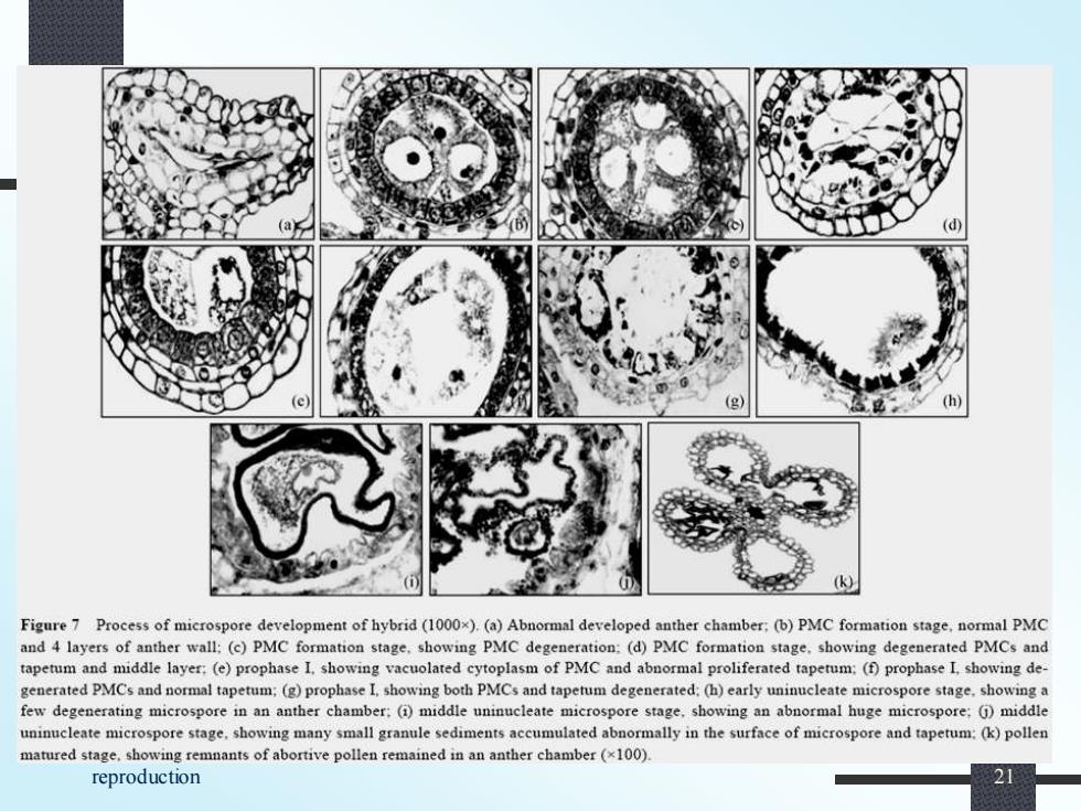
(k) Figure 7 Process of microspore development of hybrid(1000x).(a)Abnormal developed anther chamber:(b)PMC formation stage.normal PMC and 4 layers of anther wall:(c)PMC formation stage.showing PMC degeneration:(d)PMC formation stage.showing degenerated PMCs and tapetum and middle layer:(e)prophase I.showing vacuolated cytoplasm of PMC and abnormal proliferated taperum:(f)prophase I showing de- generated PMCs and normal tapetum:(g)prophase I,showing both PMCs and tapetum degenerated:(h)early uninucleate microspore stage.showing a few degenerating microspore in an anther chamber:()middle uninucleate microspore stage.showing an abnormal huge microspore:()middle uninucleate microspore stage,showing many small granule sediments accumulated abnormally in the surface of microspore and tapetum:(k)pollen matured stage.showing remnants of abortive pollen remained in an anther chamber(x100). reproduction Biology of plant reproduction 21