正在加载图片...

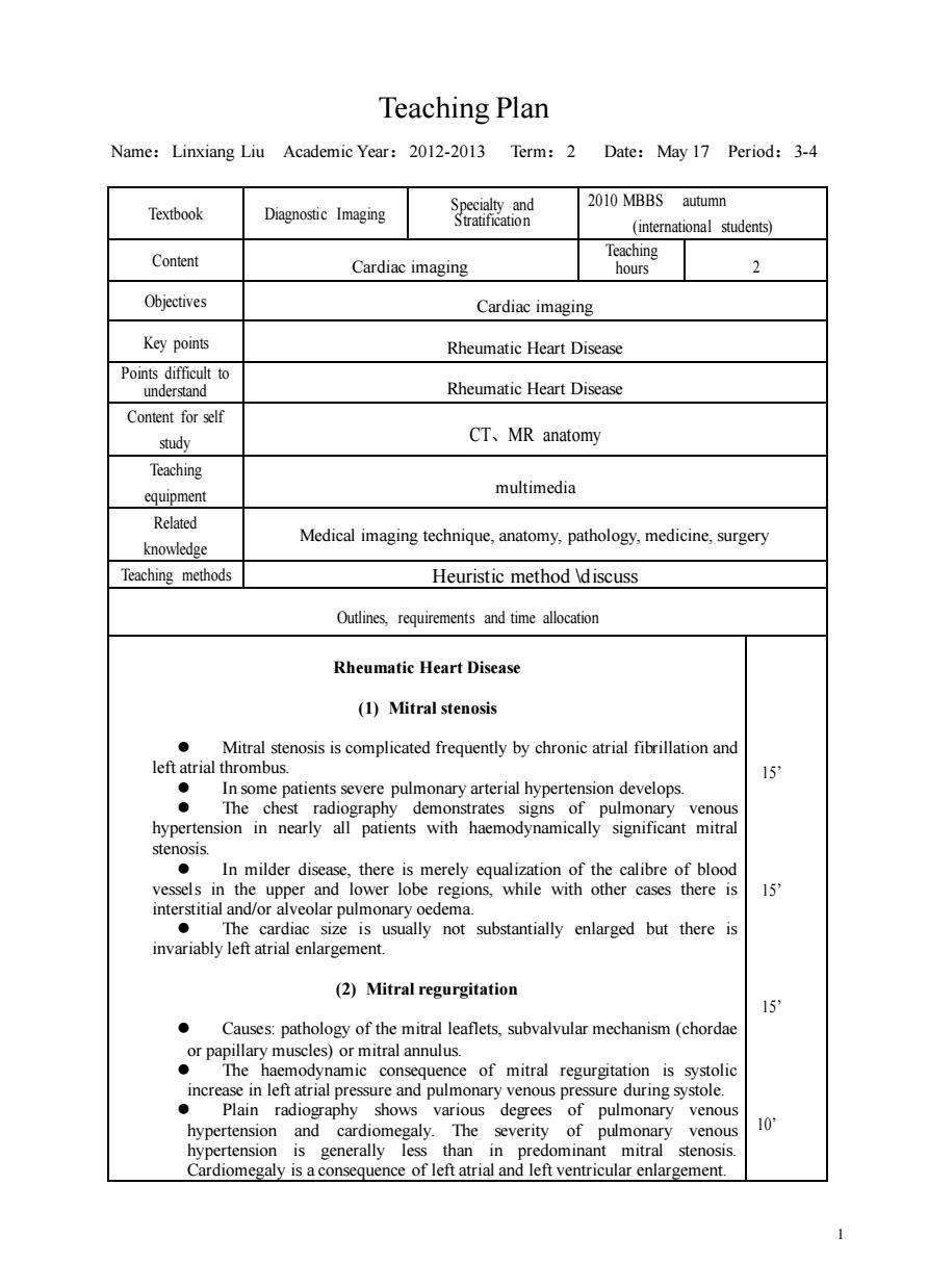
Teaching Plan Name:Linxiang Liu Academic Year:2012-2013 Term:2 Date:May 17 Period:3-4 2010 MBBS autumn Textbook Diagnostic Imaging 8e8 (students) Content Cardiac imaging Teaching 2 Objectives Cardiac imaging Key points Rheumatic Heart Disease Pointsdiffic o unders Rheumatic Heart Disease Content for self study CT、MR anatomy Teaching equipment multimedia Related knowledge Medical imaging technique,anatomy,pathology,medicine,surgery Teaching methods Heuristic method discuss Outlines,requirements and time allocation Rheumatic Heart Disease ())Mitral stenosis Mitral stenosis is complicated frequently by chronic atrial fibrillation and left atrial thrombus. In some patients severe pulmonary arterial hypertension develops. The chest pulmonary venous sten In milder diseas nualization of the calibre of blood interstitial and/or alveolar pulmonary oedema. size is usually not substantially enlarged but there is (2)Mitral regurgitation Causes:pathology of the mitral leaflets,subvalvular mechanism(chordae orpp)orrlu The monary ring sy ven hypertension and cardiomegaly.The severity of pulmonary venous 10 hypertension is generally less than in predominant mitral stenosis. Cardiomegaly is a consequence of left atrial and left ventricular enlargement. 1 Teaching Plan Name:Linxiang Liu Academic Year:2012-2013 Term:2 Date:May 17 Period:3-4 Textbook Diagnostic Imaging Specialty and Stratification 2010 MBBS autumn (international students) Content Cardiac imaging Teaching hours 2 Objectives Cardiac imaging Key points Rheumatic Heart Disease Points difficult to understand Rheumatic Heart Disease Content for self study CT、MR anatomy Teaching equipment multimedia Related knowledge Medical imaging technique, anatomy, pathology, medicine, surgery Teaching methods Heuristic method \discuss Outlines, requirements and time allocation Rheumatic Heart Disease (1) Mitral stenosis ⚫ Mitral stenosis is complicated frequently by chronic atrial fibrillation and left atrial thrombus. ⚫ In some patients severe pulmonary arterial hypertension develops. ⚫ The chest radiography demonstrates signs of pulmonary venous hypertension in nearly all patients with haemodynamically significant mitral stenosis. ⚫ In milder disease, there is merely equalization of the calibre of blood vessels in the upper and lower lobe regions, while with other cases there is interstitial and/or alveolar pulmonary oedema. ⚫ The cardiac size is usually not substantially enlarged but there is invariably left atrial enlargement. (2) Mitral regurgitation ⚫ Causes: pathology of the mitral leaflets, subvalvular mechanism (chordae or papillary muscles) or mitral annulus. ⚫ The haemodynamic consequence of mitral regurgitation is systolic increase in left atrial pressure and pulmonary venous pressure during systole. ⚫ Plain radiography shows various degrees of pulmonary venous hypertension and cardiomegaly. The severity of pulmonary venous hypertension is generally less than in predominant mitral stenosis. Cardiomegaly is a consequence of left atrial and left ventricular enlargement. 15’ 15’ 15’ 10’