正在加载图片...
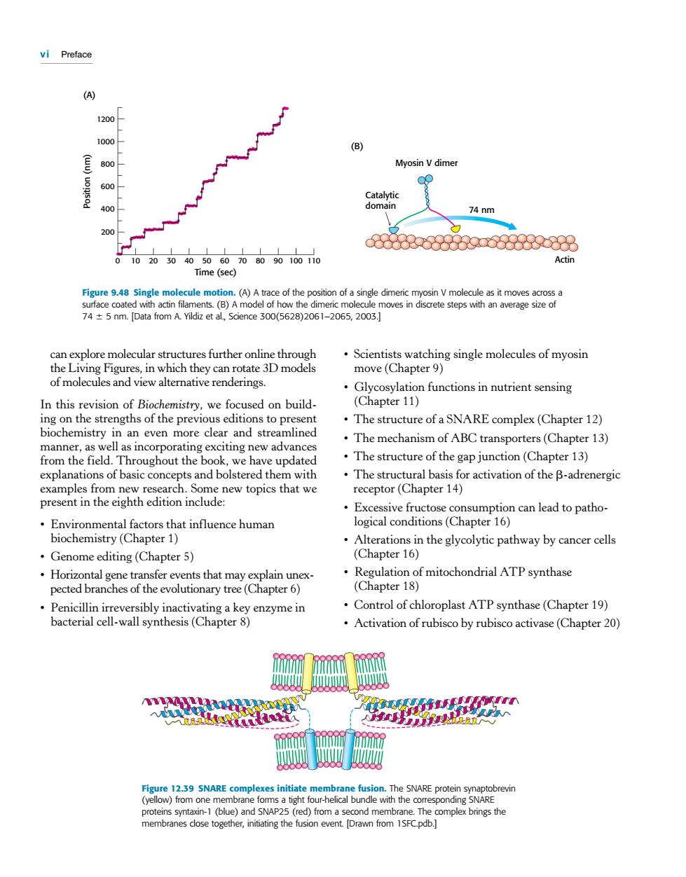
vi Preface 120oA 1000 B 800 Myosin Vdimer 40o 74 nm e300(56 the c 61-2065,2003 plore molecular str es further online thro which ate 3D models watchin of molecules and view alternative renderings Glycosylation functions in nutrient sensing In this revision of Biochemistry.we focused on build- (Chapter 11) ing on the strengths of the previous editions to present .The structure of a SNARE complex(Chapter 12) biochemistry in an even more clear and streamlined The mechanism of ABC transporters(Chapter 13) manner,as well as incorporating exciting new advance from the e field we ha update The structure of the gap junction(Chapter 13) explana ons of basic concepts and b The stru or activation of the B-adrenergic Excessive fructose consumption can lead to patho- Enviror mental factors that influence human logical conditions(Chapter 16) biochemistry (Chapter 1) .Alterations in the glycolytic pathway by cancer cells .Genome editing(Chapter 5) (Chapter 16) Horizontal gene transfer events that may explain unex pected branches of the evolutionary tree(Chapter 6) n a key Control of chloroplast ATP synthase(Chapter 19) .Activation of rubisco by rubisco activase(Chapter 20) omplex brings the vi Preface can explore molecular structures further online through the Living Figures, in which they can rotate 3D models of molecules and view alternative renderings. In this revision of Biochemistry, we focused on building on the strengths of the previous editions to present biochemistry in an even more clear and streamlined manner, as well as incorporating exciting new advances from the field. Throughout the book, we have updated explanations of basic concepts and bolstered them with examples from new research. Some new topics that we present in the eighth edition include: • Environmental factors that influence human biochemistry (Chapter 1) • Genome editing (Chapter 5) • Horizontal gene transfer events that may explain unexpected branches of the evolutionary tree (Chapter 6) • Penicillin irreversibly inactivating a key enzyme in bacterial cell-wall synthesis (Chapter 8) • Scientists watching single molecules of myosin move (Chapter 9) • Glycosylation functions in nutrient sensing (Chapter 11) • The structure of a SNARE complex (Chapter 12) • The mechanism of ABC transporters (Chapter 13) • The structure of the gap junction (Chapter 13) • The structural basis for activation of the b-adrenergic receptor (Chapter 14) • Excessive fructose consumption can lead to pathological conditions (Chapter 16) • Alterations in the glycolytic pathway by cancer cells (Chapter 16) • Regulation of mitochondrial ATP synthase (Chapter 18) • Control of chloroplast ATP synthase (Chapter 19) • Activation of rubisco by rubisco activase (Chapter 20) Time (sec) 0 10 3020 40 50 60 70 9080 100 110 Position (nm) 1200 1000 800 600 400 200 (A) Catalytic domain Myosin V dimer 74 nm Actin (B) Figure 9.48 Single molecule motion. (A) A trace of the position of a single dimeric myosin V molecule as it moves across a surface coated with actin filaments. (B) A model of how the dimeric molecule moves in discrete steps with an average size of 74 6 5 nm. [Data from A. Yildiz et al., Science 300(5628)2061–2065, 2003.] Figure 12.39 SNARE complexes initiate membrane fusion. The SNARE protein synaptobrevin (yellow) from one membrane forms a tight four-helical bundle with the corresponding SNARE proteins syntaxin-1 (blue) and SNAP25 (red) from a second membrane. The complex brings the membranes close together, initiating the fusion event. [Drawn from 1SFC.pdb.]