正在加载图片...

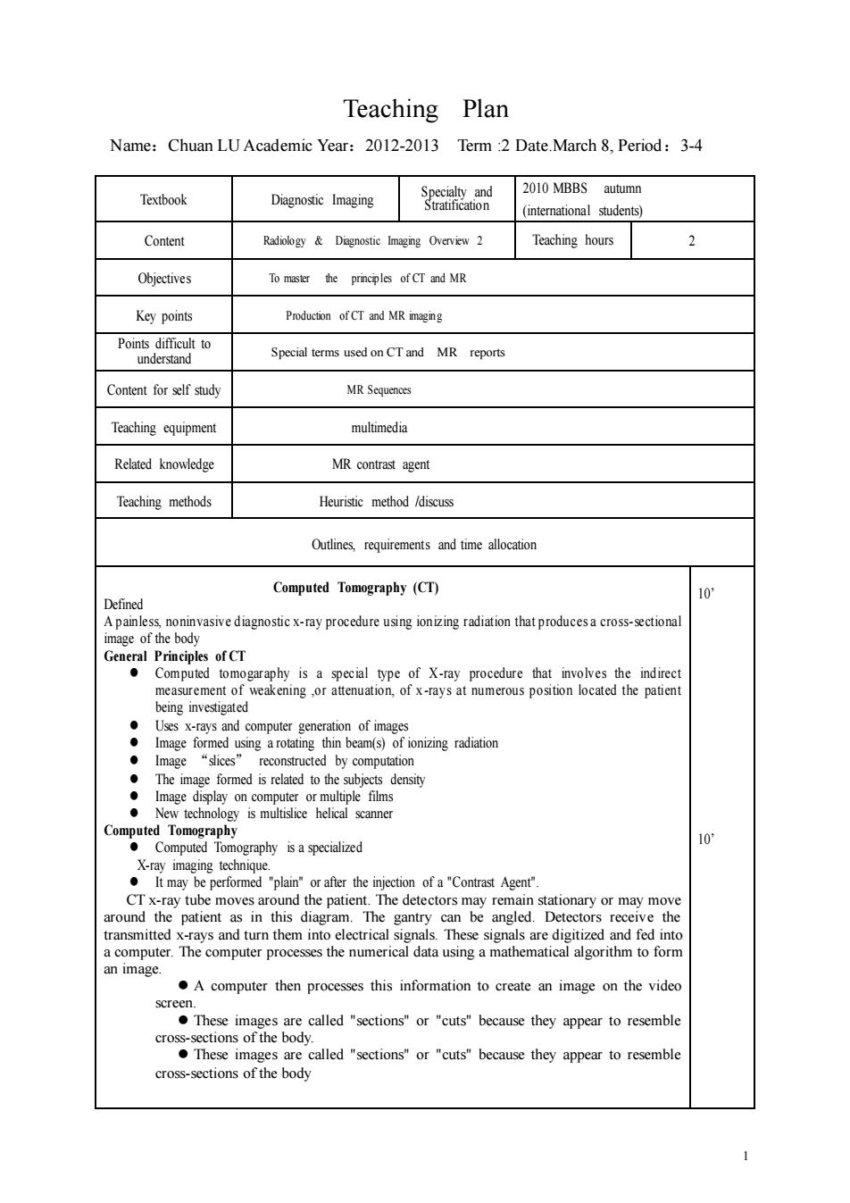
Teaching Plan Name:Chuan LU Academic Year:2012-2013 Term :2 Date.March 8,Period:3-4 2010 MBBS autumn Textbook Diagnostic Imaging a Content Teaching hours 2 Objectives To master the principles of CT ad MR Key points Production of CT and MR mgng po Special terms used onCTand MR reports Content for lf study MR Seauenos Teaching equipment multimedia Related knowedge MR contrast agent Teaching methods Heuristic method /discuss Outlines requirements and time al Computed Tomography (CT) Defined Anrksabmvasvedagnostcxayprocadireusngomzagratiaionthatpotbcesacosgdoml Geera Pripe fCT akening ,or at d usingaro beam(s)c The image formed is related to the density nography isa specialied 10 Crx-ray tube moves around the patient.The detectors may remain stationary or may move around the patient as in sdiagram.The gantry can be angled.Detector receive the an image. A computer then processes this information to create an image on the vide screen. These mere called ctionsor utsbecaurse they appear to resemble mages are called""cuts because they appear to resemble cross-sections of the body 1 1 Teaching Plan Name:Chuan LU Academic Year:2012-2013 Term :2 Date.March 8, Period:3-4 Textbook Diagnostic Imaging Specialty and Stratification 2010 MBBS autumn (international students) Content Radiology & Diagnostic Imaging Overview 2 Teaching hours 2 Objectives To master the principles of CT and MR Key points Production of CT and MR imaging Points difficult to understand Special terms used on CT and MR reports Content for self study MR Sequences Teaching equipment multimedia Related knowledge MR contrast agent Teaching methods Heuristic method /discuss Outlines, requirements and time allocation Computed Tomography (CT) Defined A painless, noninvasive diagnostic x-ray procedure using ionizing radiation that produces a cross-sectional image of the body General Principles of CT ⚫ Computed tomogaraphy is a special type of X-ray procedure that involves the indirect measurement of weakening ,or attenuation, of x -rays at numerous position located the patient being investigated ⚫ Uses x-rays and computer generation of images ⚫ Image formed using a rotating thin beam(s) of ionizing radiation ⚫ Image “slices” reconstructed by computation ⚫ The image formed is related to the subjects density ⚫ Image display on computer or multiple films ⚫ New technology is multislice helical scanner Computed Tomography ⚫ Computed Tomography is a specialized X-ray imaging technique. ⚫ It may be performed "plain" or after the injection of a "Contrast Agent". CT x-ray tube moves around the patient. The detectors may remain stationary or may move around the patient as in this diagram. The gantry can be angled. Detectors receive the transmitted x-rays and turn them into electrical signals. These signals are digitized and fed into a computer. The computer processes the numerical data using a mathematical algorithm to form an image. ⚫ A computer then processes this information to create an image on the video screen. ⚫ These images are called "sections" or "cuts" because they appear to resemble cross-sections of the body. ⚫ These images are called "sections" or "cuts" because they appear to resemble cross-sections of the body 10’ 10’