正在加载图片...

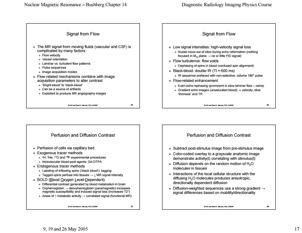
Nuclear Magnetic Resonance-Bushberg Chapter 14 Diagnostic Radiology Imaging Physics Course Signal from Flow Signal from Flow The MR signal from moving fluids (vascular and CSF)is Low signal intensities:high-velocity signal loss complicated by many factors: ation(nothing ◆Flow velocity ◆Vessel orientation .Laminar vs.turbulent flow patterns Flow turbulence:flow voids Pulse sequences Dephasing of spins in blood(confused spin alignment) Image acquistion modes Black-blood:double IR (TI=600 ms) Flow related mechanisms combine with image IR sequence prefaced with non-selective.volume 180pulse acquisition parameters to alter contrast Flow-related enhancement ."Bright-blood'to black-blood' Even-echo rephasing (prominent in slow laminar flow-veins) .Can be a source of artifacts Gradient ech Exploited to produce MR angiography images 色制 Perfusion and Diffusion Contrast Perfusion and Diffusion Contrast Perfusion of cells via capillary bed Subtract post-stimulus image from pre-stimulus image Exogenous tracer methods Color-coded overlay to a grayscale anatomic image ◆2H.3He.17oand1年experimental procedures Intravascular blood-pool agents:Gd-DTPA demonstrate activity(t)correlating with stimulus(t) Endogenous tracer methods Diffusion depends on the random motion of H2O molecules in tissues Labeling of inflowing spins (black blood"):tagging ◆Tagged spins perfuse into tissues→!MR signal intensity Interactions of the local cellular structure with the BOLD(Blood Oxygen Level-Dependent) diffusing H2O molecules produ uces anisotropic, generated by bl netabolism in brair directionally dependent diffusion Diffusion-weighted sequences use a strong gradient- Areas of metabolic activity-correlated signal (functional MR) signal differences based on mobility/directionality 9,19and26May2005 17Nuclear Magnetic Resonance – Bushberg Chapter 14 Diagnostic Radiology Imaging Physics Course 9, 19 and 26 May 2005 17 © UW and Brent K. Stewart, PhD, DABMP 65 Signal from Flow The MR signal from moving fluids (vascular and CSF) is The MR signal from moving fluids (vascular and CSF) is complicated by many factors: complicated by many factors: Flow velocity Vessel orientation Laminar vs. turbulent flow patterns Pulse sequences Image acquisition modes Flow related mechanisms combine with image Flow related mechanisms combine with image acquisition parameters to alter contrast acquisition parameters to alter contrast ‘Bright ‘Bright-blood’ to ‘black-blood’ Can be a source of artifacts Exploited to produce MR angiography images © UW and Brent K. Stewart, PhD, DABMP 66 Signal from Flow Low signal intensities: high-velocity signal loss Nuclei move out of slice during echo reformation (nothing Nuclei move out of slice during echo reformation (nothing focused in Mxy plane → no or little FID signal no or little FID signal) Flow turbulence: flow voids Dephasing of spins in blood (confused spin alignment) Dephasing of spins in blood (confused spin alignment) Black-blood: double IR (TI blood: double IR (TI ≈ 600 ms) 600 ms) IR sequence prefaced with non-selective, volume 180° pulse Flow-related enhancement related enhancement Even-echo rephasing (prominent in slow laminar flow (prominent in slow laminar flow – veins) Gradient echo images (unsaturated blood): Gradient echo images (unsaturated blood): ∝ velocity, slice velocity, slice ‘thinness’ and TR © UW and Brent K. Stewart, PhD, DABMP 67 Perfusion and Diffusion Contrast Perfusion and Diffusion Contrast Perfusion of cells via capillary bed Exogenous tracer methods 2H, 3He, 17O and 19F experimental procedures Intravascular blood-pool agents: Gd-DTPA Endogenous tracer methods Labeling of inflowing spins (‘black blood’): tagging Tagged spins perfuse into tissues Tagged spins perfuse into tissues → ↓ MR signal intensity BOLD (Blood Oxygen Level-Dependent) ependent) Differential contrast generated by blood metabolism in brain Oxyhemoglobin Oxyhemoglobin → deoxyhemoglobin (paramagnetic) increases deoxyhemoglobin (paramagnetic) increases magnetic susceptibility and induced signal loss (increased T2*) magnetic susceptibility and induced signal loss (increased T2*) Areas of Areas of ↑ metabolic activity metabolic activity → correlated signal (functional MR) correlated signal (functional MR) © UW and Brent K. Stewart, PhD, DABMP 68 Perfusion and Diffusion Contrast Perfusion and Diffusion Contrast Subtract post Subtract post-stimulus image from pre-stimulus image Color-coded overlay to a grayscale anatomic image coded overlay to a grayscale anatomic image demonstrate demonstrate activity(t activity(t) correlating with ) correlating with stimulus(t stimulus(t) Diffusion depends on the random motion of H2O molecules in tissues Interactions of the local cellular structure with the Interactions of the local cellular structure with the diffusing H2O molecules produces anisotropic, O molecules produces anisotropic, directionally dependent diffusion Diffusion-weighted sequences use a strong gradient weighted sequences use a strong gradient → signal differences based on mobility/directionality