正在加载图片...
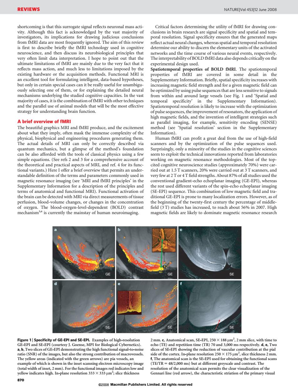
REVIEWS NATUREIVol 453112 June 2008 gate signal reflects neuronal mass acti- Critical factors determining the utility of fMRI for drawing con The aim of thi rcficetactualncural neuro ience,and then dis sneobioigcalpihapileha hypothese from within and Spatial and und Humn from the of high-fid the od's founda t of he terms oftd with MRI via direct mearements of ncentr SE SE 870 08 Macmillan Publishers Limited.All rights reservedshortcoming is that this surrogate signal reflects neuronal mass activity. Although this fact is acknowledged by the vast majority of investigators, its implications for drawing judicious conclusions from fMRI data are most frequently ignored. The aim of this review is first to describe briefly the fMRI technology used in cognitive neuroscience, and then discuss its neurobiological principles that very often limit data interpretation. I hope to point out that the ultimate limitations of fMRI are mainly due to the very fact that it reflects mass action, and much less to limitations imposed by the existing hardware or the acquisition methods. Functional MRI is an excellent tool for formulating intelligent, data-based hypotheses, but only in certain special cases can it be really useful for unambiguously selecting one of them, or for explaining the detailed neural mechanisms underlying the studied cognitive capacities. In the vast majority of cases, it is the combination of fMRI with other techniques and the parallel use of animal models that will be the most effective strategy for understanding brain function. A brief overview of fMRI The beautiful graphics MRI and fMRI produce, and the excitement about what they imply, often mask the immense complexity of the physical, biophysical and engineering procedures generating them. The actual details of MRI can only be correctly described via quantum mechanics, but a glimpse of the method’s foundation can be also afforded with the tools of classical physics using a few simple equations. (See refs 2 and 3 for a comprehensive account of the theoretical and practical aspects of MRI, and ref. 4 for its functional variants.) Here I offer a brief overview that permits an understandable definition of the terms and parameters commonly used in magnetic resonance imaging (see ‘MRI and fMRI principles’ in the Supplementary Information for a description of the principles and terms of anatomical and functional MRI). Functional activation of the brain can be detected with MRI via direct measurements of tissue perfusion, blood-volume changes, or changes in the concentration of oxygen. The blood-oxygen-level-dependent (BOLD) contrast mechanism5,6 is currently the mainstay of human neuroimaging. Critical factors determining the utility of fMRI for drawing conclusions in brain research are signal specificity and spatial and temporal resolution. Signal specificity ensures that the generated maps reflect actual neural changes, whereas spatial and temporal resolution determine our ability to discern the elementary units of the activated networks and the time course of various neural events, respectively. The interpretability of BOLD fMRI data also depends critically on the experimental design used. Spatiotemporal properties of BOLD fMRI. The spatiotemporal properties of fMRI are covered in some detail in the Supplementary Information. Briefly, spatial specificity increases with increasing magnetic field strength and for a given magnetic field can be optimized by using pulse sequences that are less sensitive to signals from within and around large vessels (see Fig. 1 and ‘Spatial and temporal specificity’ in the Supplementary Information). Spatiotemporal resolution is likely to increase with the optimization of pulse sequences, the improvement of resonators, the application of high magnetic fields, and the invention of intelligent strategies such as parallel imaging, for example, sensitivity encoding (SENSE) method (see ‘Spatial resolution’ section in the Supplementary Information). Human fMRI can profit a great deal from the use of high-field scanners and by the optimization of the pulse sequences used. Surprisingly, only a minority of the studies in the cognitive sciences seem to exploit the technical innovations reported from laboratories working on magnetic resonance methodologies. Most of the topcited cognitive neuroscience studies (approximately 70%) were carried out at 1.5 T scanners, 20% were carried out at 3 T scanners, and very few at 2 T or 4 T field strengths. About 87% of all studies used the conventional gradient-echo echoplanar imaging (GE-EPI), whereas the rest used different variants of the spin-echo echoplanar imaging (SE-EPI) sequence. This combination of low magnetic field and traditional GE-EPI is prone to many localization errors. However, as of the beginning of the twenty-first century the percentage of middlefield (3 T) studies has increased, to reach about 56% in 2007. High magnetic fields are likely to dominate magnetic resonance research a bc d ef GE GE SE SE SE SE Figure 1 | Specificity of GE-EPI and SE-EPI. Examples of high-resolution GE-EPI and SE-EPI (courtesy J. Goense, MPI for Biological Cybernetics). a, b, Two slices of GE-EPI demonstrating the high functional signal-to-noise ratio (SNR) of the images, but also the strong contribution of macrovessels. The yellow areas (indicated with the green arrows) are pia vessels, an example of which is shown in the inset scanning electron microscopy image (total width of inset, 2 mm). For the functional images red indicates low and yellow indicates high. In-plane resolution 333 3 333 mm2 ; slice thickness 2 mm. c, Anatomical scan, SE-EPI, 250 3 188 mm2 , 2 mm slice, with time to echo (TE) and repetition time (TR) 70 and 3,000 ms respectively. d, e, Two slices of SE-EPI showing the reduction of vascular contribution at the pial side of the cortex. In-plane resolution 250 3 175 mm2 , slice thickness 2 mm. f, The anatomical scan is the SE-EPI used for obtaining the functional scans (TE/TR 5 48/2,000 ms) but at different greyscale and contrast. The resolution of the anatomical scan permits the clear visualization of the Gennari line (red arrow), the characteristic striation of the primary visual REVIEWS NATUREjVol 453j12 June 2008 870 ©2008 Macmillan Publishers Limited. All rights reserved