正在加载图片...
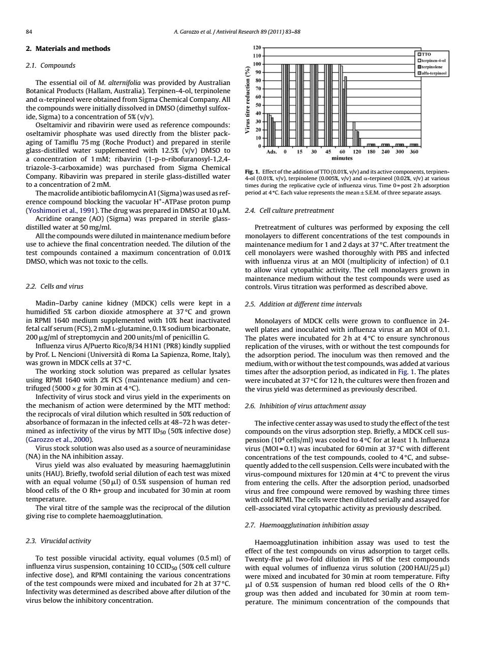
84 A Garozzo et aL Antiviral Research 89(2011)83-88 2.Materials and methods 120 110 口TT0 terpinen-4-o 2.1.Compounds 100 terpinolene 90 口ala-4 erpineol The essential oil of M.alternifolia was provided by Australian 80 Botanical Products(Hallam,Australia).Terpinen-4-ol,terpinolene 70 and o-terpineol were obtained from Sigma Chemical Company.All 60 the compounds were initially dissolved in DMSO(dimethyl sulfox- ide,Sigma)to a concentration of 5%(v/v). Oseltamivir and ribavirin were used as reference compounds: oseltamivir phosphate was used directly from the blister pack- aging of Tamiflu 75mg(Roche Product)and prepared in sterile glass-distilled water supplemented with 12.5%(v/v)DMSO to Ads 0 15 30 4560120180240300360 a concentration of 1 mM;ribavirin (1-p-D-ribofuranosyl-1,2,4- minutes triazole-3-carboxamide)was purchased from Sigma Chemical Fig.1.Effect of the addition of TTO(0.01%,v/v)and its active components,terpinen- Company.Ribavirin was prepared in sterile glass-distilled water 4-ol (0.01%.v/v).terpinolene (0.005%.v/v)and a-terpineol (0.02%.v/v)at various to a concentration of 2 mM. times during the replicative cycle of influenza virus.Time 0=post 2 h adsorption The macrolide antibiotic bafilomycin A1(Sigma)was used as ref- period at 4C.Each value represents the mean+S.E.M.of three separate assays. erence compound blocking the vacuolar H*-ATPase proton pump (Yoshimori et al.,1991).The drug was prepared in DMSO at 10 uM 2.4.Cell culture pretreatment Acridine orange (AO)(Sigma)was prepared in sterile glass- distilled water at 50 mg/ml. Pretreatment of cultures was performed by exposing the cell All the compounds were diluted in maintenance medium before monolayers to different concentrations of the test compounds in use to achieve the final concentration needed.The dilution of the maintenance medium for 1 and 2 days at 37C.After treatment the test compounds contained a maximum concentration of 0.01% cell monolayers were washed thoroughly with PBS and infected DMSO.which was not toxic to the cells. with influenza virus at an MOI(multiplicity of infection)of 0.1 to allow viral cytopathic activity.The cell monolayers grown in maintenance medium without the test compounds were used as 2.2.Cells and virus controls.Virus titration was performed as described above. Madin-Darby canine kidney (MDCK)cells were kept in a 2.5.Addition at different time intervals humidified 5%carbon dioxide atmosphere at 37C and grown in RPMI 1640 medium supplemented with 10%heat inactivated Monolayers of MDCK cells were grown to confluence in 24- fetal calf serum(FCS).2 mM L-glutamine,0.1%sodium bicarbonate, well plates and inoculated with influenza virus at an MOl of 0.1 200 wg/ml of streptomycin and 200 units/ml of penicillin G. The plates were incubated for 2h at 4C to ensure synchronous Influenza virus A/Puerto Rico/8/34 H1N1 (PR8)kindly supplied replication of the viruses,with or without the test compounds for by Prof.L Nencioni (Universita di Roma La Sapienza,Rome,Italy). the adsorption period.The inoculum was then removed and the was grown in MDCK cells at 37C. medium,with or without the test compounds,was added at various The working stock solution was prepared as cellular lysates times after the adsorption period,as indicated in Fig.1.The plates using RPMI 1640 with 2%FCS (maintenance medium)and cen- were incubated at 37C for 12 h,the cultures were then frozen and trifuged (5000 x g for 30 min at 4C). the virus yield was determined as previously described. Infectivity of virus stock and virus yield in the experiments on the mechanism of action were determined by the MTT method: 2.6.Inhibition of virus attachment assay the reciprocals of viral dilution which resulted in 50%reduction of absorbance of formazan in the infected cells at 48-72 h was deter- The infective center assay was used to study the effect of the test mined as infectivity of the virus by MTT IDs0(50%infective dose) compounds on the virus adsorption step.Briefly,a MDCK cell sus- (Garozzo et al.,2000). pension (104 cells/ml)was cooled to 4C for at least 1 h.Influenza Virus stock solution was also used as a source of neuraminidase virus(MOI=0.1)was incubated for 60min at 37C with different (NA)in the NA inhibition assay. concentrations of the test compounds,cooled to 4C,and subse- Virus yield was also evaluated by measuring haemagglutinin quently added to the cell suspension.Cells were incubated with the units (HAU).Briefly,twofold serial dilution of each test was mixed virus-compound mixtures for 120 min at 4C to prevent the virus with an equal volume (50 ul)of 0.5%suspension of human red from entering the cells.After the adsorption period,unadsorbed blood cells of the O Rh+group and incubated for 30 min at room virus and free compound were removed by washing three times temperature. with cold RPMI.The cells were then diluted serially and assayed for The viral titre of the sample was the reciprocal of the dilution cell-associated viral cytopathic activity as previously described. giving rise to complete haemoagglutination. 2.7.Haemoagglutination inhibition assay 2.3.Virucidal activity Haemoagglutination inhibition assay was used to test the effect of the test compounds on virus adsorption to target cells. To test possible virucidal activity.equal volumes(0.5 ml)of Twenty-five ul two-fold dilution in PBS of the test compounds influenza virus suspension,containing 10 CCIDso(50%cell culture with equal volumes of influenza virus solution (200 HAU/25 wl) infective dose).and RPMI containing the various concentrations were mixed and incubated for 30 min at room temperature.Fifty of the test compounds were mixed and incubated for 2h at 37C. ul of 0.5%suspension of human red blood cells of the O Rh+ Infectivity was determined as described above after dilution of the group was then added and incubated for 30min at room tem- virus below the inhibitory concentration. perature.The minimum concentration of the compounds that84 A. Garozzo et al. / Antiviral Research 89 (2011) 83–88 2. Materials and methods 2.1. Compounds The essential oil of M. alternifolia was provided by Australian Botanical Products (Hallam, Australia). Terpinen-4-ol, terpinolene and -terpineol were obtained from Sigma Chemical Company. All the compounds were initially dissolved in DMSO (dimethyl sulfoxide, Sigma) to a concentration of 5% (v/v). Oseltamivir and ribavirin were used as reference compounds: oseltamivir phosphate was used directly from the blister packaging of Tamiflu 75 mg (Roche Product) and prepared in sterile glass-distilled water supplemented with 12.5% (v/v) DMSO to a concentration of 1 mM; ribavirin (1-p-d-ribofuranosyl-1,2,4- triazole-3-carboxamide) was purchased from Sigma Chemical Company. Ribavirin was prepared in sterile glass-distilled water to a concentration of 2 mM. Themacrolide antibiotic bafilomycin A1 (Sigma) was used as reference compound blocking the vacuolar H+-ATPase proton pump (Yoshimori et al., 1991). The drug was prepared in DMSO at 10 M. Acridine orange (AO) (Sigma) was prepared in sterile glassdistilled water at 50 mg/ml. All the compounds were diluted in maintenance medium before use to achieve the final concentration needed. The dilution of the test compounds contained a maximum concentration of 0.01% DMSO, which was not toxic to the cells. 2.2. Cells and virus Madin–Darby canine kidney (MDCK) cells were kept in a humidified 5% carbon dioxide atmosphere at 37 ◦C and grown in RPMI 1640 medium supplemented with 10% heat inactivated fetal calf serum (FCS), 2 mM l-glutamine, 0.1% sodium bicarbonate, 200 g/ml of streptomycin and 200 units/ml of penicillin G. Influenza virus A/Puerto Rico/8/34 H1N1 (PR8) kindly supplied by Prof. L. Nencioni (Università di Roma La Sapienza, Rome, Italy), was grown in MDCK cells at 37 ◦C. The working stock solution was prepared as cellular lysates using RPMI 1640 with 2% FCS (maintenance medium) and centrifuged (5000 × g for 30 min at 4 ◦C). Infectivity of virus stock and virus yield in the experiments on the mechanism of action were determined by the MTT method: the reciprocals of viral dilution which resulted in 50% reduction of absorbance of formazan in the infected cells at 48–72 h was determined as infectivity of the virus by MTT ID50 (50% infective dose) (Garozzo et al., 2000). Virus stock solution was also used as a source of neuraminidase (NA) in the NA inhibition assay. Virus yield was also evaluated by measuring haemagglutinin units (HAU). Briefly, twofold serial dilution of each test was mixed with an equal volume (50 l) of 0.5% suspension of human red blood cells of the O Rh+ group and incubated for 30 min at room temperature. The viral titre of the sample was the reciprocal of the dilution giving rise to complete haemoagglutination. 2.3. Virucidal activity To test possible virucidal activity, equal volumes (0.5 ml) of influenza virus suspension, containing 10 CCID50 (50% cell culture infective dose), and RPMI containing the various concentrations of the test compounds were mixed and incubated for 2 h at 37 ◦C. Infectivity was determined as described above after dilution of the virus below the inhibitory concentration. Fig. 1. Effect of the addition of TTO (0.01%, v/v) and its active components, terpinen- 4-ol (0.01%, v/v), terpinolene (0.005%, v/v) and -terpineol (0.02%, v/v) at various times during the replicative cycle of influenza virus. Time 0 = post 2 h adsorption period at 4 ◦C. Each value represents the mean ± S.E.M. of three separate assays. 2.4. Cell culture pretreatment Pretreatment of cultures was performed by exposing the cell monolayers to different concentrations of the test compounds in maintenance medium for 1 and 2 days at 37 ◦C. After treatment the cell monolayers were washed thoroughly with PBS and infected with influenza virus at an MOI (multiplicity of infection) of 0.1 to allow viral cytopathic activity. The cell monolayers grown in maintenance medium without the test compounds were used as controls. Virus titration was performed as described above. 2.5. Addition at different time intervals Monolayers of MDCK cells were grown to confluence in 24- well plates and inoculated with influenza virus at an MOI of 0.1. The plates were incubated for 2 h at 4 ◦C to ensure synchronous replication of the viruses, with or without the test compounds for the adsorption period. The inoculum was then removed and the medium, with or without the test compounds, was added at various times after the adsorption period, as indicated in Fig. 1. The plates were incubated at 37 ◦C for 12 h, the cultures were then frozen and the virus yield was determined as previously described. 2.6. Inhibition of virus attachment assay The infective center assay was used to study the effect of the test compounds on the virus adsorption step. Briefly, a MDCK cell suspension (104 cells/ml) was cooled to 4 ◦C for at least 1 h. Influenza virus (MOI = 0.1) was incubated for 60 min at 37 ◦C with different concentrations of the test compounds, cooled to 4 ◦C, and subsequently added to the cell suspension. Cells were incubated with the virus-compound mixtures for 120 min at 4 ◦C to prevent the virus from entering the cells. After the adsorption period, unadsorbed virus and free compound were removed by washing three times with cold RPMI. The cells were then diluted serially and assayed for cell-associated viral cytopathic activity as previously described. 2.7. Haemoagglutination inhibition assay Haemoagglutination inhibition assay was used to test the effect of the test compounds on virus adsorption to target cells. Twenty-five l two-fold dilution in PBS of the test compounds with equal volumes of influenza virus solution (200 HAU/25 l) were mixed and incubated for 30 min at room temperature. Fifty l of 0.5% suspension of human red blood cells of the O Rh+ group was then added and incubated for 30 min at room temperature. The minimum concentration of the compounds that��������