正在加载图片...
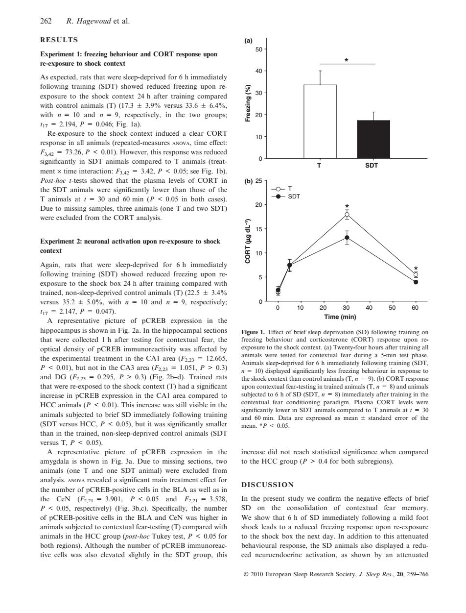
262 R.Hagewoud et al. RESULTS (a) 50 Experiment 1:freezing behaviour and CORT response upon re-exposure to shock context 40 As expected,rats that were sleep-deprived for 6 h immediately following training (SDT)showed reduced freezing upon re- 8 exposure to the shock context 24 h after training compared 30 with control animals(T)(17.3±3.9%versus33.6±6.4%, with n=10 and n=9,respectively,in the two groups; 20 117=2.194,P=0.046:Fig.1a). Re-exposure to the shock context induced a clear CORT 10 response in all animals (repeated-measures ANOvA,time effect: F3.42 =73.26,P <0.01).However,this response was reduced significantly in SDT animals compared to T animals (treat- 0 SDT ment x time interaction:F3.42 =3.42,P<0.05;see Fig.Ib). Post-hoc t-tests showed that the plasma levels of CORT in (b)251 the SDT animals were significantly lower than those of the -O-T T animals at t 30 and 60 min (P<0.05 in both cases). ◆SDT Due to missing samples,three animals (one T and two SDT) 20 ★ were excluded from the CORT analysis. 15 Experiment 2:neuronal activation upon re-exposure to shock 骂 context 10 Again,rats that were sleep-deprived for 6 h immediately following training (SDT)showed reduced freezing upon re- 5 exposure to the shock box 24 h after training compared with ● trained,non-sleep-deprived control animals(T)(22.5+3.4% versus 35.2+5.0%,with n 10 and n =9,respectively; 117=2.147,P=0.047. 10 2030 40 50 鸟 A representative picture of pCREB expression in the Time(min) hippocampus is shown in Fig.2a.In the hippocampal sections Figure 1.Effect of brief sleep deprivation(SD)following training on that were collected I h after testing for contextual fear,the freezing behaviour and corticosterone (CORT)response upon re- optical density of pCREB immunoreactivity was affected by exposure to the shock context.(a)Twenty-four hours after training all the experimental treatment in the CAl area(F223 12.665, animals were tested for contextual fear during a 5-min test phase. Animals sleep-deprived for 6 h immediately following training(SDT. P<0.01),but not in the CA3 area (F2.23 1.051,P>0.3) n=10)displayed significantly less freezing behaviour in response to and DG (F2.23 =0.295,P>0.3)(Fig.2b-d).Trained rats the shock context than control animals (T,n 9).(b)CORT response that were re-exposed to the shock context (T)had a significant upon contextual fear-testing in trained animals (T,n=8)and animals increase in pCREB expression in the CAl area compared to subjected to 6 h of SD(SDT,n=8)immediately after training in the HCC animals (P<0.01).This increase was still visible in the contextual fear conditioning paradigm.Plasma CORT levels were animals subjected to brief SD immediately following training significantly lower in SDT animals compared to T animals at t=30 and 60 min.Data are expressed as mean standard error of the (SDT versus HCC,P 0.05),but it was significantly smaller mean.*P 0.05 than in the trained,non-sleep-deprived control animals(SDT versus T,P 0.05). A representative picture of pCREB expression in the increase did not reach statistical significance when compared amygdala is shown in Fig.3a.Due to missing sections,two to the HCC group (P>0.4 for both subregions). animals (one T and one SDT animal)were excluded from analysis.ANOvA revealed a significant main treatment effect for DISCUSSION the number of pCREB-positive cells in the BLA as well as in the CeN(F2.21=3.901,P<0.05andf2.21=3.528, In the present study we confirm the negative effects of brief P 0.05,respectively)(Fig.3b,c).Specifically,the number SD on the consolidation of contextual fear memory. of pCREB-positive cells in the BLA and CeN was higher in We show that 6 h of SD immediately following a mild foot animals subjected to contextual fear-testing (T)compared with shock leads to a reduced freezing response upon re-exposure animals in the HCC group (post-hoc Tukey test,P<0.05 for to the shock box the next day.In addition to this attenuated both regions).Although the number of pCREB immunoreac- behavioural response,the SD animals also displayed a redu- tive cells was also elevated slightly in the SDT group,this ced neuroendocrine activation,as shown by an attenuated 2010 European Sleep Research Society,J.Sleep Res..20,259-266RESULTS Experiment 1: freezing behaviour and CORT response upon re-exposure to shock context As expected, rats that were sleep-deprived for 6 h immediately following training (SDT) showed reduced freezing upon reexposure to the shock context 24 h after training compared with control animals (T) (17.3 ± 3.9% versus 33.6 ± 6.4%, with n = 10 and n = 9, respectively, in the two groups; t17 = 2.194, P = 0.046; Fig. 1a). Re-exposure to the shock context induced a clear CORT response in all animals (repeated-measures anova, time effect: F3,42 = 73.26, P < 0.01). However, this response was reduced significantly in SDT animals compared to T animals (treatment · time interaction: F3,42 = 3.42, P < 0.05; see Fig. 1b). Post-hoc t-tests showed that the plasma levels of CORT in the SDT animals were significantly lower than those of the T animals at t = 30 and 60 min (P < 0.05 in both cases). Due to missing samples, three animals (one T and two SDT) were excluded from the CORT analysis. Experiment 2: neuronal activation upon re-exposure to shock context Again, rats that were sleep-deprived for 6 h immediately following training (SDT) showed reduced freezing upon reexposure to the shock box 24 h after training compared with trained, non-sleep-deprived control animals (T) (22.5 ± 3.4% versus 35.2 ± 5.0%, with n = 10 and n = 9, respectively; t17 = 2.147, P = 0.047). A representative picture of pCREB expression in the hippocampus is shown in Fig. 2a. In the hippocampal sections that were collected 1 h after testing for contextual fear, the optical density of pCREB immunoreactivity was affected by the experimental treatment in the CA1 area (F2,23 = 12.665, P < 0.01), but not in the CA3 area (F2,23 = 1.051, P > 0.3) and DG (F2,23 = 0.295, P > 0.3) (Fig. 2b–d). Trained rats that were re-exposed to the shock context (T) had a significant increase in pCREB expression in the CA1 area compared to HCC animals (P < 0.01). This increase was still visible in the animals subjected to brief SD immediately following training (SDT versus HCC, P < 0.05), but it was significantly smaller than in the trained, non-sleep-deprived control animals (SDT versus T, P < 0.05). A representative picture of pCREB expression in the amygdala is shown in Fig. 3a. Due to missing sections, two animals (one T and one SDT animal) were excluded from analysis. anova revealed a significant main treatment effect for the number of pCREB-positive cells in the BLA as well as in the CeN (F2,21 = 3.901, P < 0.05 and F2,21 = 3.528, P < 0.05, respectively) (Fig. 3b,c). Specifically, the number of pCREB-positive cells in the BLA and CeN was higher in animals subjected to contextual fear-testing (T) compared with animals in the HCC group (post-hoc Tukey test, P < 0.05 for both regions). Although the number of pCREB immunoreactive cells was also elevated slightly in the SDT group, this increase did not reach statistical significance when compared to the HCC group (P > 0.4 for both subregions). DISCUSSION In the present study we confirm the negative effects of brief SD on the consolidation of contextual fear memory. We show that 6 h of SD immediately following a mild foot shock leads to a reduced freezing response upon re-exposure to the shock box the next day. In addition to this attenuated behavioural response, the SD animals also displayed a reduced neuroendocrine activation, as shown by an attenuated T SDT Freezing (%) 0 10 20 30 40 50 * Time (min) 0 10 20 30 40 50 60 CORT (µg dL–1) 0 5 10 15 20 25 T SDT * * (a) (b) Figure 1. Effect of brief sleep deprivation (SD) following training on freezing behaviour and corticosterone (CORT) response upon reexposure to the shock context. (a) Twenty-four hours after training all animals were tested for contextual fear during a 5-min test phase. Animals sleep-deprived for 6 h immediately following training (SDT, n = 10) displayed significantly less freezing behaviour in response to the shock context than control animals (T, n = 9). (b) CORT response upon contextual fear-testing in trained animals (T, n = 8) and animals subjected to 6 h of SD (SDT, n = 8) immediately after training in the contextual fear conditioning paradigm. Plasma CORT levels were significantly lower in SDT animals compared to T animals at t = 30 and 60 min. Data are expressed as mean ± standard error of the mean. *P < 0.05. 262 R. Hagewoud et al. 2010 European Sleep Research Society, J. Sleep Res., 20, 259–266�