正在加载图片...
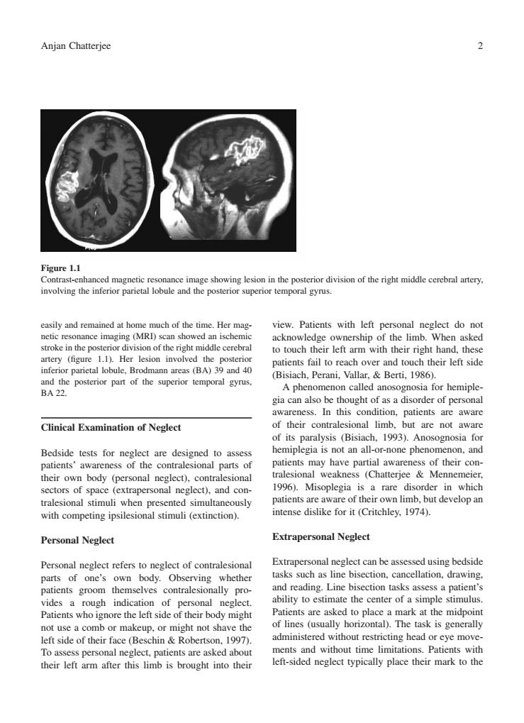
Anjan Chatterjee 2 eshowing esion in the posterior division of the right middle erera artery involving the inferior parietal lobule and the posterior superior temporal gyrus. easily and remained at hon much of the time.Her mg ership lim artery (figure 1.1).Her lesion involved the posterio inferior parietal lobule,Brodmann areas (BA)39 and 40 h over and touch their left sid poerir pat of the uerior a for hemipl gia can also be thought of as a disorder of personal awareness. In this condition.patients are aware Clinical Examination of Neglect of their contralesional limb,but are not aware of its paralysis (Bisiach,1993).Anosognosia for Bedside tests for neglect are designed to asses hemiplegia is not an all-or-none phenomenon,and atients' awareness of the contralesional parts of patients may have partial awareness of their con- their own body (personal ntralesiona tralesional weakness (Chatterjee Mennemeier sectors of space eglect)and cor 1996).Misoplegia is a rare disorder in which tralesional s simulta patients are aware of their own limb,but develop an with competing ipsilesional stimuli(extinction) intense dislike for it (Critchley.1974). Personal Neglect Extrapersonal Neglect Personal neglect refers to neglect of contralesiona Extrapersonal neglect can be assessed using bedside parts wn tasks such as line bisection.cancellation,drawing dy. ents groor ally pro and reading.Line bisection tasks assess a patient's ability to estimate the center of a simple stimulus Patients wh perso left sideo their body migh Patients are asked to place a mark at the midpoint of lines (usually horizontal).The task is generally e a c or m eup,or might not shave the left side of their face(Beschin Robertson, 99 administered without restricting head or eye move- To assess personal neglect,patients are asked about ments and without time limitations.Patients with their left arm after this limb is brought into their left-sided neglect typically place their mark to the easily and remained at home much of the time. Her magnetic resonance imaging (MRI) scan showed an ischemic stroke in the posterior division of the right middle cerebral artery (figure 1.1). Her lesion involved the posterior inferior parietal lobule, Brodmann areas (BA) 39 and 40 and the posterior part of the superior temporal gyrus, BA 22. Clinical Examination of Neglect Bedside tests for neglect are designed to assess patients’ awareness of the contralesional parts of their own body (personal neglect), contralesional sectors of space (extrapersonal neglect), and contralesional stimuli when presented simultaneously with competing ipsilesional stimuli (extinction). Personal Neglect Personal neglect refers to neglect of contralesional parts of one’s own body. Observing whether patients groom themselves contralesionally provides a rough indication of personal neglect. Patients who ignore the left side of their body might not use a comb or makeup, or might not shave the left side of their face (Beschin & Robertson, 1997). To assess personal neglect, patients are asked about their left arm after this limb is brought into their view. Patients with left personal neglect do not acknowledge ownership of the limb. When asked to touch their left arm with their right hand, these patients fail to reach over and touch their left side (Bisiach, Perani, Vallar, & Berti, 1986). A phenomenon called anosognosia for hemiplegia can also be thought of as a disorder of personal awareness. In this condition, patients are aware of their contralesional limb, but are not aware of its paralysis (Bisiach, 1993). Anosognosia for hemiplegia is not an all-or-none phenomenon, and patients may have partial awareness of their contralesional weakness (Chatterjee & Mennemeier, 1996). Misoplegia is a rare disorder in which patients are aware of their own limb, but develop an intense dislike for it (Critchley, 1974). Extrapersonal Neglect Extrapersonal neglect can be assessed using bedside tasks such as line bisection, cancellation, drawing, and reading. Line bisection tasks assess a patient’s ability to estimate the center of a simple stimulus. Patients are asked to place a mark at the midpoint of lines (usually horizontal). The task is generally administered without restricting head or eye movements and without time limitations. Patients with left-sided neglect typically place their mark to the Anjan Chatterjee 2 Figure 1.1 Contrast-enhanced magnetic resonance image showing lesion in the posterior division of the right middle cerebral artery, involving the inferior parietal lobule and the posterior superior temporal gyrus