正在加载图片...
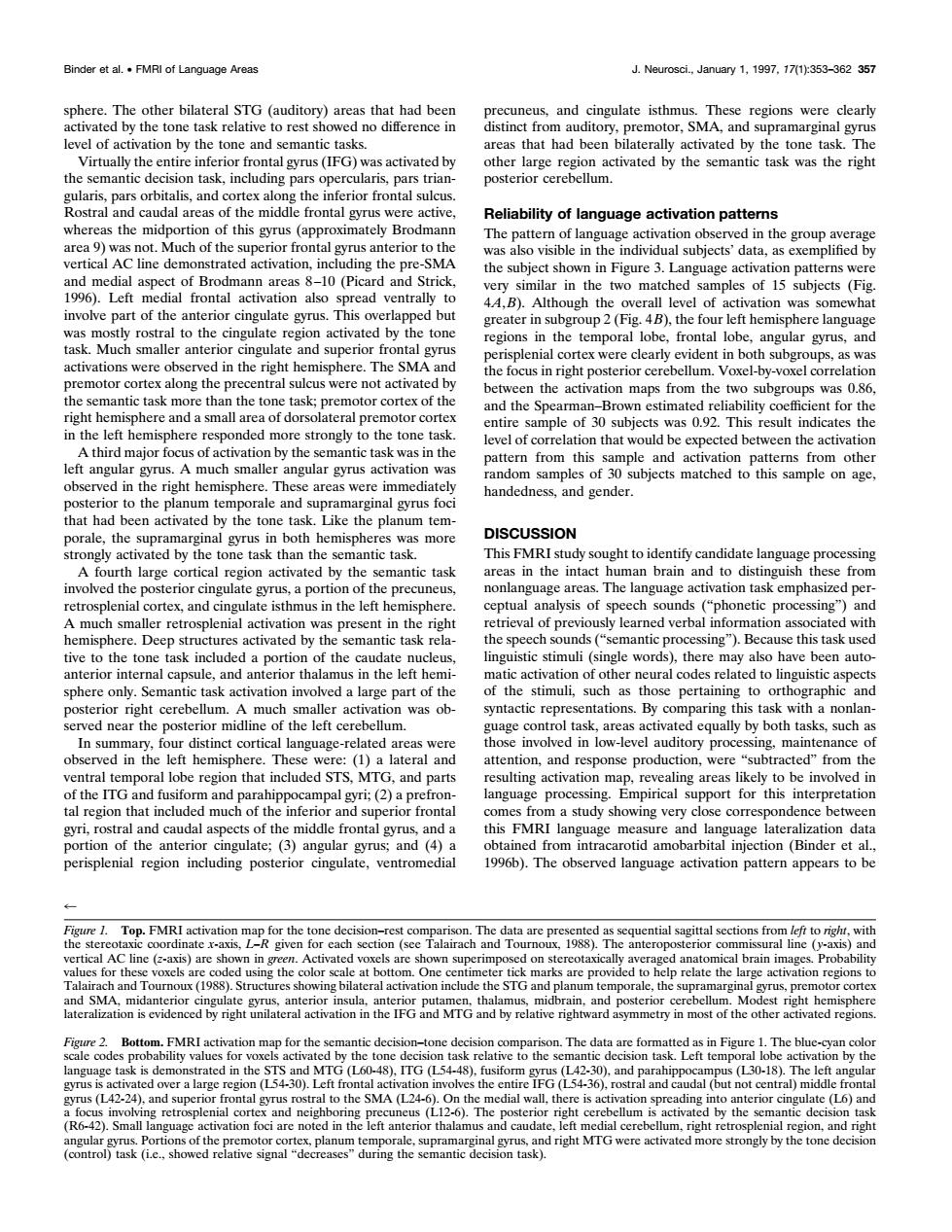
Binder eal.·MRl of Language Area Neurosci.,January 1.1997.17(1k353-362 35 level of activation by the tone and semantic tasks. lariiparsotbitalsandcortexaloathcntcriorfontalslcu5 Reliability of language activation pattems and media tiond and strick. and the level of correlation that would be expected between the activatior much angular gyrus activatior handedness,and gender hat had Like the pla num tem DISCUSSION activated by the anti cortex,and isph tive to the tone task incl a portio the cau inguistic stimuli(single words),there may also have been auto of the stimuli.such as those pertaining to orthog aphie and erior right cerebe mu sma ler activation was ob- .By comparing this task with a nonlar ve-rela ted areas we ing,maint nd ubtra form and par anguage proces inte atio n tha astudy owing very cl dence his FMri la dat angul y and (4) d from ntrac region ng po Talairach .1988 ng the m.On clate the nd Tou a The bl ask is de cd in the tkativ to the semantis d por and MTG p)The let right (1 driehtMTGWeeactivatedmoretongtheonedesphere. The other bilateral STG (auditory) areas that had been activated by the tone task relative to rest showed no difference in level of activation by the tone and semantic tasks. Virtually the entire inferior frontal gyrus (IFG) was activated by the semantic decision task, including pars opercularis, pars triangularis, pars orbitalis, and cortex along the inferior frontal sulcus. Rostral and caudal areas of the middle frontal gyrus were active, whereas the midportion of this gyrus (approximately Brodmann area 9) was not. Much of the superior frontal gyrus anterior to the vertical AC line demonstrated activation, including the pre-SMA and medial aspect of Brodmann areas 8 –10 (Picard and Strick, 1996). Left medial frontal activation also spread ventrally to involve part of the anterior cingulate gyrus. This overlapped but was mostly rostral to the cingulate region activated by the tone task. Much smaller anterior cingulate and superior frontal gyrus activations were observed in the right hemisphere. The SMA and premotor cortex along the precentral sulcus were not activated by the semantic task more than the tone task; premotor cortex of the right hemisphere and a small area of dorsolateral premotor cortex in the left hemisphere responded more strongly to the tone task. A third major focus of activation by the semantic task was in the left angular gyrus. A much smaller angular gyrus activation was observed in the right hemisphere. These areas were immediately posterior to the planum temporale and supramarginal gyrus foci that had been activated by the tone task. Like the planum temporale, the supramarginal gyrus in both hemispheres was more strongly activated by the tone task than the semantic task. A fourth large cortical region activated by the semantic task involved the posterior cingulate gyrus, a portion of the precuneus, retrosplenial cortex, and cingulate isthmus in the left hemisphere. A much smaller retrosplenial activation was present in the right hemisphere. Deep structures activated by the semantic task relative to the tone task included a portion of the caudate nucleus, anterior internal capsule, and anterior thalamus in the left hemisphere only. Semantic task activation involved a large part of the posterior right cerebellum. A much smaller activation was observed near the posterior midline of the left cerebellum. In summary, four distinct cortical language-related areas were observed in the left hemisphere. These were: (1) a lateral and ventral temporal lobe region that included STS, MTG, and parts of the ITG and fusiform and parahippocampal gyri; (2) a prefrontal region that included much of the inferior and superior frontal gyri, rostral and caudal aspects of the middle frontal gyrus, and a portion of the anterior cingulate; (3) angular gyrus; and (4) a perisplenial region including posterior cingulate, ventromedial precuneus, and cingulate isthmus. These regions were clearly distinct from auditory, premotor, SMA, and supramarginal gyrus areas that had been bilaterally activated by the tone task. The other large region activated by the semantic task was the right posterior cerebellum. Reliability of language activation patterns The pattern of language activation observed in the group average was also visible in the individual subjects’ data, as exemplified by the subject shown in Figure 3. Language activation patterns were very similar in the two matched samples of 15 subjects (Fig. 4A,B). Although the overall level of activation was somewhat greater in subgroup 2 (Fig. 4B), the four left hemisphere language regions in the temporal lobe, frontal lobe, angular gyrus, and perisplenial cortex were clearly evident in both subgroups, as was the focus in right posterior cerebellum. Voxel-by-voxel correlation between the activation maps from the two subgroups was 0.86, and the Spearman–Brown estimated reliability coefficient for the entire sample of 30 subjects was 0.92. This result indicates the level of correlation that would be expected between the activation pattern from this sample and activation patterns from other random samples of 30 subjects matched to this sample on age, handedness, and gender. DISCUSSION This FMRI study sought to identify candidate language processing areas in the intact human brain and to distinguish these from nonlanguage areas. The language activation task emphasized perceptual analysis of speech sounds (“phonetic processing”) and retrieval of previously learned verbal information associated with the speech sounds (“semantic processing”). Because this task used linguistic stimuli (single words), there may also have been automatic activation of other neural codes related to linguistic aspects of the stimuli, such as those pertaining to orthographic and syntactic representations. By comparing this task with a nonlanguage control task, areas activated equally by both tasks, such as those involved in low-level auditory processing, maintenance of attention, and response production, were “subtracted” from the resulting activation map, revealing areas likely to be involved in language processing. Empirical support for this interpretation comes from a study showing very close correspondence between this FMRI language measure and language lateralization data obtained from intracarotid amobarbital injection (Binder et al., 1996b). The observed language activation pattern appears to be 4 Figure 1. Top. FMRI activation map for the tone decision–rest comparison. The data are presented as sequential sagittal sections from left to right, with the stereotaxic coordinate x-axis, L–R given for each section (see Talairach and Tournoux, 1988). The anteroposterior commissural line ( y-axis) and vertical AC line (z-axis) are shown in green. Activated voxels are shown superimposed on stereotaxically averaged anatomical brain images. Probability values for these voxels are coded using the color scale at bottom. One centimeter tick marks are provided to help relate the large activation regions to Talairach and Tournoux (1988). Structures showing bilateral activation include the STG and planum temporale, the supramarginal gyrus, premotor cortex and SMA, midanterior cingulate gyrus, anterior insula, anterior putamen, thalamus, midbrain, and posterior cerebellum. Modest right hemisphere lateralization is evidenced by right unilateral activation in the IFG and MTG and by relative rightward asymmetry in most of the other activated regions. Figure 2. Bottom. FMRI activation map for the semantic decision–tone decision comparison. The data are formatted as in Figure 1. The blue-cyan color scale codes probability values for voxels activated by the tone decision task relative to the semantic decision task. Left temporal lobe activation by the language task is demonstrated in the STS and MTG (L60-48), ITG (L54-48), fusiform gyrus (L42-30), and parahippocampus (L30-18). The left angular gyrus is activated over a large region (L54-30). Left frontal activation involves the entire IFG (L54-36), rostral and caudal (but not central) middle frontal gyrus (L42-24), and superior frontal gyrus rostral to the SMA (L24-6). On the medial wall, there is activation spreading into anterior cingulate (L6) and a focus involving retrosplenial cortex and neighboring precuneus (L12-6). The posterior right cerebellum is activated by the semantic decision task (R6-42). Small language activation foci are noted in the left anterior thalamus and caudate, left medial cerebellum, right retrosplenial region, and right angular gyrus. Portions of the premotor cortex, planum temporale, supramarginal gyrus, and right MTG were activated more strongly by the tone decision (control) task (i.e., showed relative signal “decreases” during the semantic decision task). Binder et al. • FMRI of Language Areas J. Neurosci., January 1, 1997, 17(1):353–362 357