正在加载图片...
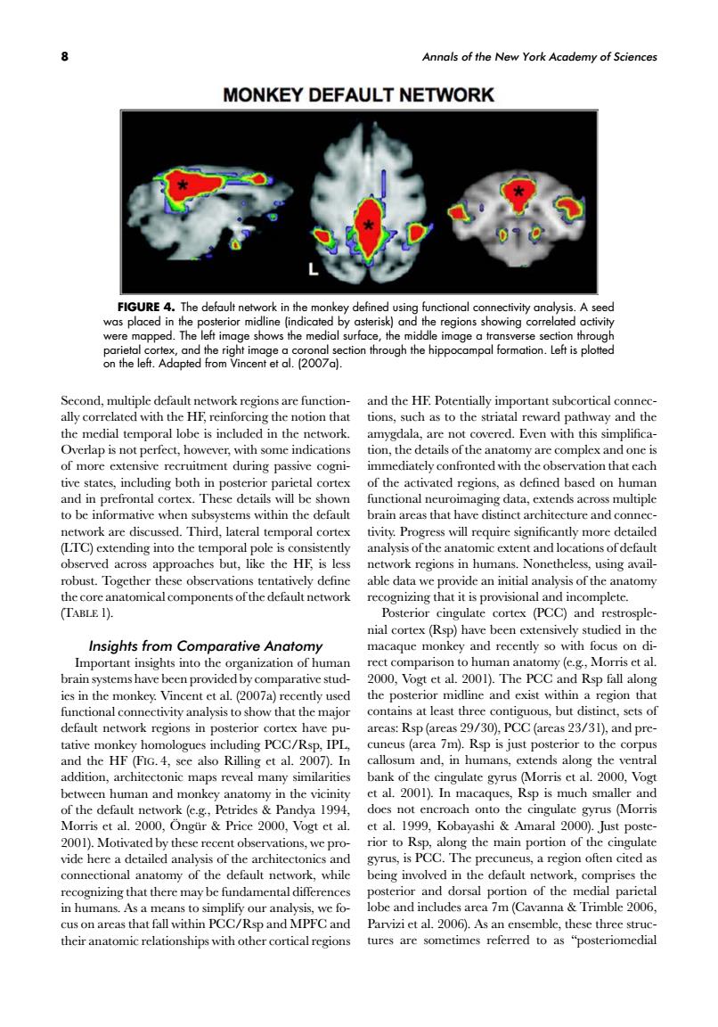
Annals of the New York Academy of Sciences MONKEY DEFAULT NETWORK was placed d.The ddleegionssho ving correla 9 Se nd the morant subcortical connce way an p0 th of more extensivr recruitment during passive tive states,including both in posterior parietal cortex of the activated regions,as defined based on human and in prefrontal c ortex.These details will be showr functional neuroimasing data.extends across multiple to be informative when subsystems within the default brain areas that have distinct architecture and conne ork are Third,lateral HE is lc an robust.Tog e obse able dat n initial analysis of the that it is provisional and incomplete (TABLE 1). Posterior cingulate cortex (PCC)and restrosple- nial cortex(Rsp)have been extensively studied in the Insights from Comparative Anatomy macaque monkey and recently so with focus on di man an my (c.g.Mor 0. 1 nat least three contig default network regions in po ureas:Rsp(areas29/30).PCC(areas 23/31).and pre- tative monkey homologues including PCC/Rsp,IPL cuneus (area 7m).Rsp is just posterior to the corpus and the HF (FIG.4,see also Rilling et al.2007).In callosum and,in humans,extends along the ventral addition,architectonic maps reveal many similarities nthe vicinity et al.2001).In caques, s.Pric s(Mo 00 v shi 2001)Motivated by the the main gyrus,is PCC.The precuneus,a region often cited as connectional anatomy of the default network.while being involved in the default network,comprises the recognizing that there may be fundamental differences C/ we lo and i ludes are a 7m (Cavanna cus on areas tha Rsp and Parvizi et al. thesc threest 8 Annals of the New York Academy of Sciences FIGURE 4. The default network in the monkey defined using functional connectivity analysis. A seed was placed in the posterior midline (indicated by asterisk) and the regions showing correlated activity were mapped. The left image shows the medial surface, the middle image a transverse section through parietal cortex, and the right image a coronal section through the hippocampal formation. Left is plotted on the left. Adapted from Vincent et al. (2007a). Second, multiple default network regions are functionally correlated with the HF, reinforcing the notion that the medial temporal lobe is included in the network. Overlap is not perfect, however, with some indications of more extensive recruitment during passive cognitive states, including both in posterior parietal cortex and in prefrontal cortex. These details will be shown to be informative when subsystems within the default network are discussed. Third, lateral temporal cortex (LTC) extending into the temporal pole is consistently observed across approaches but, like the HF, is less robust. Together these observations tentatively define the core anatomical components of the default network (TABLE 1). Insights from Comparative Anatomy Important insights into the organization of human brain systems have been provided by comparative studies in the monkey. Vincent et al. (2007a) recently used functional connectivity analysis to show that the major default network regions in posterior cortex have putative monkey homologues including PCC/Rsp, IPL, and the HF (FIG. 4, see also Rilling et al. 2007). In addition, architectonic maps reveal many similarities between human and monkey anatomy in the vicinity of the default network (e.g., Petrides & Pandya 1994, Morris et al. 2000, Ong ¨ ur¨ & Price 2000, Vogt et al. 2001). Motivated by these recent observations, we provide here a detailed analysis of the architectonics and connectional anatomy of the default network, while recognizing that there may be fundamental differences in humans. As a means to simplify our analysis, we focus on areas that fall within PCC/Rsp and MPFC and their anatomic relationships with other cortical regions and the HF. Potentially important subcortical connections, such as to the striatal reward pathway and the amygdala, are not covered. Even with this simplification, the details of the anatomy are complex and one is immediately confronted with the observation that each of the activated regions, as defined based on human functional neuroimaging data, extends across multiple brain areas that have distinct architecture and connectivity. Progress will require significantly more detailed analysis of the anatomic extent and locations of default network regions in humans. Nonetheless, using available data we provide an initial analysis of the anatomy recognizing that it is provisional and incomplete. Posterior cingulate cortex (PCC) and restrosplenial cortex (Rsp) have been extensively studied in the macaque monkey and recently so with focus on direct comparison to human anatomy (e.g., Morris et al. 2000, Vogt et al. 2001). The PCC and Rsp fall along the posterior midline and exist within a region that contains at least three contiguous, but distinct, sets of areas: Rsp (areas 29/30), PCC (areas 23/31), and precuneus (area 7m). Rsp is just posterior to the corpus callosum and, in humans, extends along the ventral bank of the cingulate gyrus (Morris et al. 2000, Vogt et al. 2001). In macaques, Rsp is much smaller and does not encroach onto the cingulate gyrus (Morris et al. 1999, Kobayashi & Amaral 2000). Just posterior to Rsp, along the main portion of the cingulate gyrus, is PCC. The precuneus, a region often cited as being involved in the default network, comprises the posterior and dorsal portion of the medial parietal lobe and includes area 7m (Cavanna & Trimble 2006, Parvizi et al. 2006). As an ensemble, these three structures are sometimes referred to as “posteriomedial