正在加载图片...
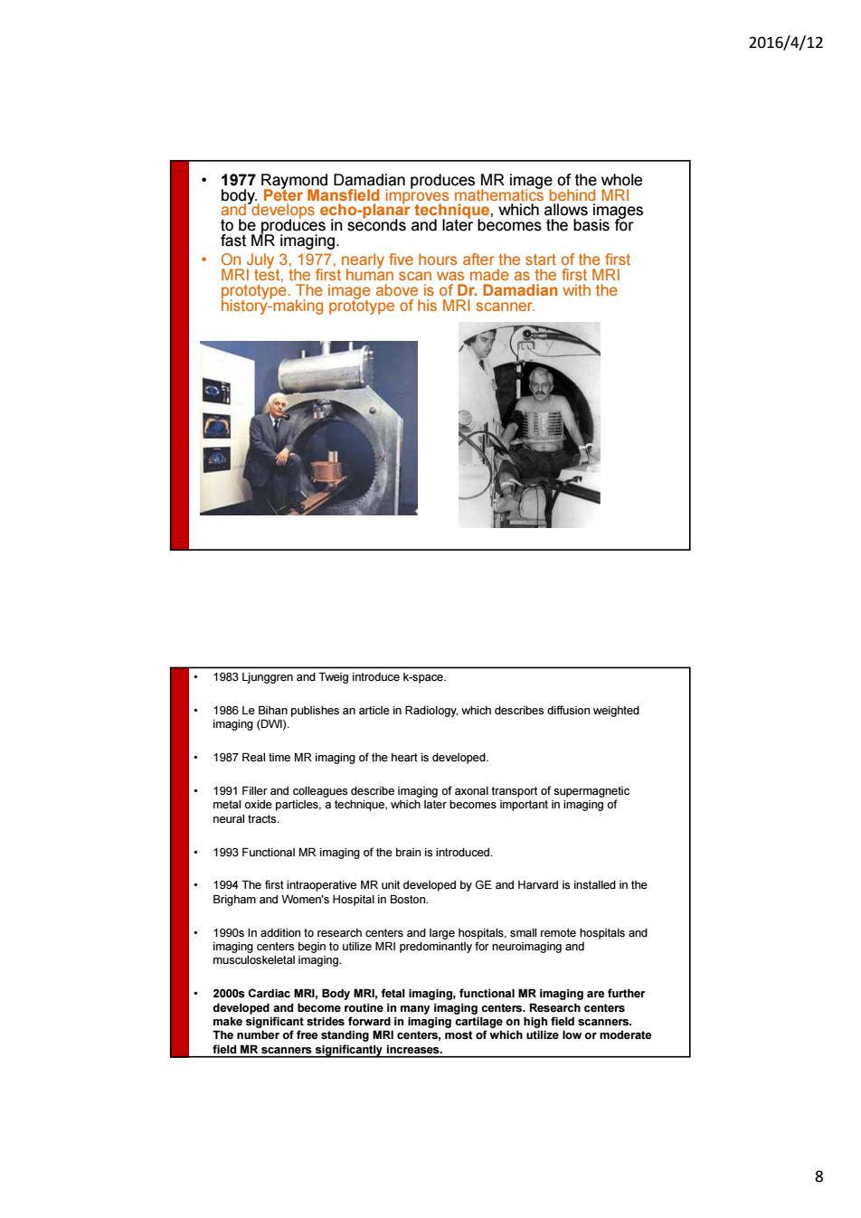
2016/4/12 ech ar tec which allo 319 nearly five hours after the start of the first dinwith the 1987Real time MR imaging of the heart is developed. neural tracts of the brain is introduced. 1990s In addition to of f g MRI enters, MR nners significantly increase 8 2016/4/12 8 • 1977 Raymond Damadian produces MR image of the whole body. Peter Mansfield improves mathematics behind MRI and develops echo-planar technique, which allows images to be produces in seconds and later becomes the basis for fast MR imaging. • On July 3, 1977, nearly five hours after the start of the first MRI test, the first human scan was made as the first MRI prototype. The image above is of Dr. Damadian with the history-making prototype of his MRI scanner. • 1983 Ljunggren and Tweig introduce k-space. • 1986 Le Bihan publishes an article in Radiology, which describes diffusion weighted imaging (DWI). • 1987 Real time MR imaging of the heart is developed. • 1991 Filler and colleagues describe imaging of axonal transport of supermagnetic metal oxide particles, a technique, which later becomes important in imaging of neural tracts. • 1993 Functional MR imaging of the brain is introduced. • 1994 The first intraoperative MR unit developed by GE and Harvard is installed in the Brigham and Women's Hospital in Boston. • 1990s In addition to research centers and large hospitals, small remote hospitals and imaging centers begin to utilize MRI predominantly for neuroimaging and musculoskeletal imaging. • 2000s Cardiac MRI, Body MRI, fetal imaging, functional MR imaging are further developed and become routine in many imaging centers. Research centers make significant strides forward in imaging cartilage on high field scanners. The number of free standing MRI centers, most of which utilize low or moderate field MR scanners significantly increases