正在加载图片...
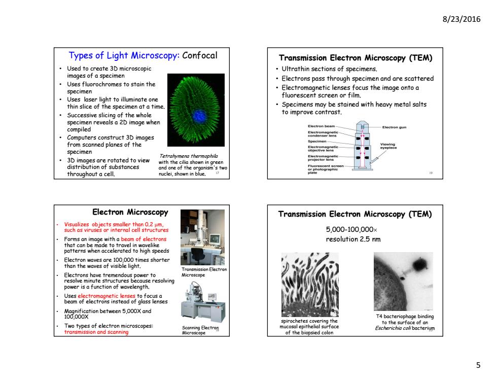
8/23/2016 Types of Light Microscopy:Confocal Transmission Electron Microscopy (TEM) Used to create 3D microscopic Ultrathin sections of specimens. images of a specimen Electrons pass through specimen and are scattered Uses fluorochromes to stain the specimen Electromagnetic lenses focus the image onto a Uses laser light to illuminate one fluorescent screen or film. thin slice of the specimen at a time Specimens may be stained with heavy metal salts Successive slicing of the whole to improve contrast. specimen reveals a 2D image when Electron beam compiled Computers construct 3D images from scanned planes of the specimen Tetrahymena thermnoohila rotated to view with the cilia shown in green F8for8na折 substances .sho and one o f the throughout a cell. sop Electron Microscopy Transmission Electron Microscopy (TEM) hr地g92es 5.000-100.000¥ resolution 2.5 nm patterns when accelerated to high speeds ro power is a function of wavelength. 638般oabetnmen5i0oxand rochetes cover ing the T4 bacteriophage binding Tw types of el fron microscopes osal epithelic of the 5 8/23/2016 5 • Used to create 3D microscopic images of a specimen • Uses fluorochromes to stain the specimen • Uses laser light to illuminate one thin slice of the specimen at a time. • Successive slicing of the whole specimen reveals a 2D image when compiled • Computers construct 3D images from scanned planes of the specimen • 3D images are rotated to view distribution of substances throughout a cell. Types of Light Microscopy: Confocal Tetrahymena thermophila with the cilia shown in green and one of the organism's two nuclei, shown in blue. 17 Electron Microscopy • Visualizes objects smaller than 0.2 m, such as viruses or internal cell structures • Forms an image with a beam of electrons that can be made to travel in wavelike patterns when accelerated to high speeds • Electron waves are 100,000 times shorter than the waves of visible light. • Electrons have tremendous power to resolve minute structures because resolving power is a function of wavelength. • Uses electromagnetic lenses to focus a beam of electrons instead of glass lenses • Magnification between 5,000X and 100,000X • Two types of electron microscopes: transmission and scanning Transmission Electron Microscope Scanning Electron Microscope 18 • Ultrathin sections of specimens. • Electrons pass through specimen and are scattered • Electromagnetic lenses focus the image onto a fluorescent screen or film. • Specimens may be stained with heavy metal salts to improve contrast. Transmission Electron Microscopy (TEM) 19 5,000-100,000 resolution 2.5 nm Transmission Electron Microscopy (TEM) T4 bacteriophage binding to the surface of an Escherichia coli bacterium spirochetes covering the mucosal epithelial surface of the biopsied colon 20