正在加载图片...
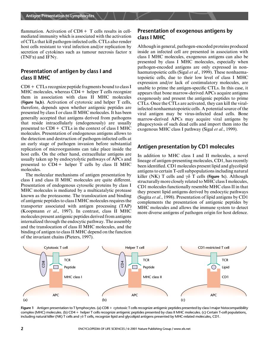
Antigen Presentation to Lymphocytes Presentation of exogenous antigens by of CTLs that kill patho class I MHC fAotceisTeistaRaooialiniectionandorreplicationby Although in general,pathogen-encoded proteins produced nted h ssed in non Presentation of antigen by class I and class ll MHC topoietic cells 伦m-c appears that bone marrow-derived APCs acquire antigens therefore,depends upon whether antigenic pe otides are the CTLs are activ ated,they can presented by class I or class II MHC molecules.It has been marrow-derived APCs may acquire viral antigens by oge ntia Antigen presentation by CD1 molecules h usually taken up by endocytolytic pathways of aPCs and isens to certain T-cellsubpopulations including natural killer (NK)T cells and y T cells (Figure 1c).Although sentation of endogenous cytosolic proteins by class I structurally more closely related to MH MHC molecules is mediated by a multicatalytic protease I mol e M tha ().Presentation of lipid antigens by CD pep complements the presentation of antigenic peptides by vith Koopmamn1In sonua cMHC MHC molecules a and allow edthrough ereptdes derivedom n end on the functior of the invariant chains(Pieters,1997). CD1-restricted Tcel Pep MHC class APC (a) (C) n toT lympho ding natural r (N ( Is and yo recognize lipid and glycolipid antigens present ed by Mh ted mol 2 ENCYCLOPEDIA OF LIFE SCIENCES/2001 Nature Publishing Group /www.els.net flammation. Activation of CD8 1 T cells results in cellmediated immunity which is associated with the activation of CTLs that kill pathogen-infected cells. CTLs also render host cells resistant to viral infection and/or replication by secretion of cytokines such as tumour necrosis factor a (TNFa) and IFNg. Presentation of antigen by class I and class II MHC CD8 1 CTLs recognize peptide fragments bound to class I MHC molecules, whereas CD4 1 helper T cells recognize them in association with class II MHC molecules (Figure 1a,b). Activation of cytotoxic and helper T cells, therefore, depends upon whether antigenic peptides are presented by class I or class II MHC molecules. It has been generally accepted that antigens derived from pathogens that reside intracellularly (endogenously) are usually presented to CD8 1 CTLs in the context of class I MHC molecules. Presentation of endogenous antigens allows to the detection and destruction of pathogen-infected cells at an early stage of pathogen invasion before substantial replication of microorganisms can take place inside the host cells. On the other hand, extracellular antigens are usually taken up by endocytolytic pathways of APCs and presented to CD4 1 helper T cells by class II MHC molecules. The molecular mechanisms of antigen presentation by class I and class II MHC molecules are quite different. Presentation of endogenous cytosolic proteins by class I MHC molecules is mediated by a multicatalytic protease known as the proteasome. The translocation and binding of antigenic peptides to class IMHC molecules requires the transporter associated with antigen processing (TAP) (Koopmann et al., 1997). In contrast, class II MHC molecules present antigenic peptides derived from antigens internalized through the endocytic pathway. The assembly and the translocation of class II MHC molecules, and the binding of antigen to class II MHC depend on the function of the invariant chains (Pieters, 1997). Presentation of exogenous antigens by class I MHC Although in general, pathogen-encoded proteins produced inside an infected cell are presented in association with class I MHC molecules, exogenous antigens can also be presented by class I MHC molecules, especially when pathogen-encoded antigens are only expressed in nonhaematopoietic cells (Sigal et al., 1999). These nonhaematopoietic cells, due to their low level of class I MHC expression and/or lack of costimulatory molecules, are unable to prime the antigen-specific CTLs. In this case, it appears that bone marrow-derived APCs acquire antigens exogenously and present the antigenic peptides to prime CTLs. Once the CTLs are activated, they can kill the viralinfected nonhaematopoietic cells. A potential source of the viral antigen may be virus-infected dead cells. Bone marrow-derived APCs may acquire viral antigens by phagocytosis of such dead cells and import them into the exogenous MHC class I pathway (Sigal et al., 1999). Antigen presentation by CD1 molecules In addition to MHC class I and II molecules, a novel lineage of antigen-presenting molecules, CD1, has recently been identified. CD1 molecules present lipid and glycolipid antigens to certain T-cell subpopulations including natural killer (NK) T cells and gd T cells (Figure 1c). Although structurally more closely related toMHC class I molecules, CD1 molecules functionally resemble MHC class II in that they present lipid antigens derived by endocytic pathways (Sugita et al., 1998). Presentation of lipid antigens by CD1 complements the presentation of antigenic peptides by MHC molecules and allows the immune system to detect more diverse antigens of pathogen origin for host defence. Cytotoxic T cell (a) (b) (c) Helper T cell CD1-restricted T cell APC APC APC Peptide TCR CD8 MHC class I Peptide TCR CD4 MHC class II Lipid TCR CD1 Figure 1 Antigen presentation to T lymphocytes. (a) CD8 1 cytotoxic T cells recognize antigenic peptides presented by class I major histocompatibility complex (MHC) molecules. (b) CD4 1 helper T cells recognize antigenic peptides presented by class II MHC molecules. (c) Certain T-cell populations, including natural killer (NK) T cells and gd T cells, recognize lipid and glycolipid antigens presented by MHC-related molecules, CD1. Antigen Presentation to Lymphocytes 2 ENCYCLOPEDIA OF LIFE SCIENCES / & 2001 Nature Publishing Group / www.els.net