正在加载图片...
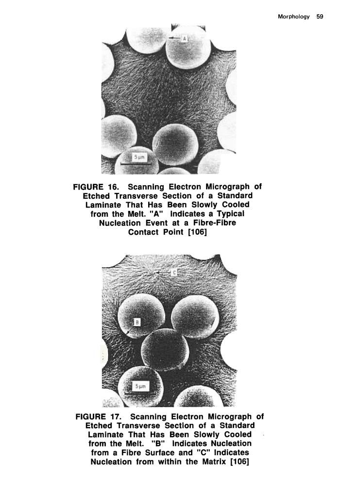
Morphology 59 5 FIGURE 16.Scanning Electron Micrograph of Etched Transverse Section of a Standard Laminate That Has Been Slowly Cooled from the Melt."A"Indicates a Typical Nucleation Event at a Fibre-Fibre Contact Point [106] 名 FIGURE 17.Scanning Electron Micrograph of Etched Transverse Section of a Standard Laminate That Has Been Slowly Cooled from the Melt."B"Indicates Nucleation from a Fibre Surface and "C"Indicates Nucleation from within the Matrix [106]Morphology 59 FIGURE 16. Scanning Electron Micrograph of Etched Transverse Section of a Standard Laminate That Has Been Slowly Cooled from the Melt." A " Indicates a Typical Nucleation Event at a Fibre-Fibre Contact Point [106] FIGURE 17. Scanning Electron Micrograph of Etched Transverse Section of a Standard Laminate That Has Been Slowly Cooled from the Melt. IIB'1 Indicates Nucleation from a Fibre Surface and "C" Indicates Nucleation from within the Matrix [106]