正在加载图片...
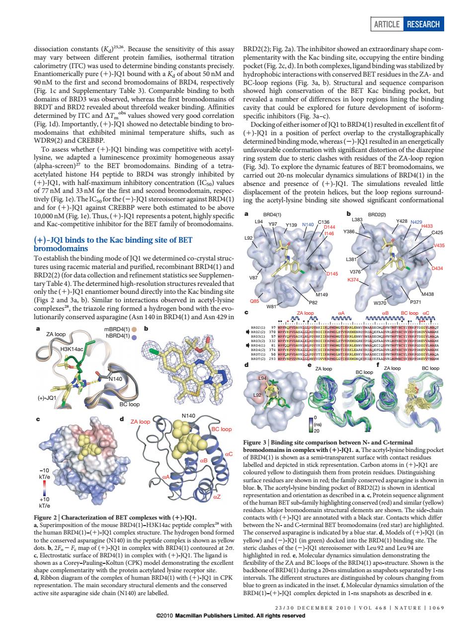
ARTICLE RESEARCH D2(2):Fi T ty with Fig. hle hin he nun lues sho ed very go correlation cavity that could be explored for future development ofisinding R9(2)and CRI ding mode,w hereas()-Q1 resul ed ir with assa ring em due to steric s of the ZA-loop region lated his H4 P peptide to BRD4 was stro f BET of BRD4()in the ce and mce of (+)-JQ1. The ation revealed litt :1 Y139 (nds to the Kac bindingsite of BET Toestablish the binding mode of determined co-crystal stru and b).Si in a lutionarily conserved asparagine (Asn 140 in BRD40D)and Asn 429n and C-termina ms in (+)-JQI a t of BRD2(2)is shown in id lina.c.F onof BET with (+)-JQ Q1 ar of( (+)-1O1.Ths li ement with the p 2010 Macdissociation constants (Kd) 25,26. Because the sensitivity of this assay may vary between different protein families, isothermal titration calorimetry (ITC) was used to determine binding constants precisely. Enantiomerically pure (1)-JQ1 bound with a Kd of about 50 nM and 90 nM to the first and second bromodomains of BRD4, respectively (Fig. 1c and Supplementary Table 3). Comparable binding to both domains of BRD3 was observed, whereas the first bromodomains of BRDT and BRD2 revealed about threefold weaker binding. Affinities determined by ITC and DTm obs values showed very good correlation (Fig. 1d). Importantly, (1)-JQ1 showed no detectable binding to bromodomains that exhibited minimal temperature shifts, such as WDR9(2) and CREBBP. To assess whether (1)-JQ1 binding was competitive with acetyllysine, we adapted a luminescence proximity homogeneous assay (alpha-screen)27 to the BET bromodomains. Binding of a tetraacetylated histone H4 peptide to BRD4 was strongly inhibited by (1)-JQ1, with half-maximum inhibitory concentration (IC50) values of 77 nM and 33 nM for the first and second bromodomain, respectively (Fig. 1e). The IC50 for the (2)-JQ1 stereoisomer against BRD4(1) and for (1)-JQ1 against CREBBP were both estimated to be above 10,000 nM (Fig. 1e). Thus, (1)-JQ1 represents a potent, highly specific and Kac-competitive inhibitor for the BET family of bromodomains. (1)-JQ1 binds to the Kac binding site of BET bromodomains To establish the binding mode of JQ1 we determined co-crystal structures using racemic material and purified, recombinant BRD4(1) and BRD2(2) (for data collection and refinement statistics see Supplementary Table 4). The determined high-resolution structures revealed that only the (1)-JQ1 enantiomer bound directly into the Kac binding site (Figs 2 and 3a, b). Similar to interactions observed in acetyl-lysine complexes28, the triazole ring formed a hydrogen bond with the evolutionarily conserved asparagine (Asn 140 in BRD4(1) and Asn 429 in BRD2(2); Fig. 2a). The inhibitor showed an extraordinary shape complementarity with the Kac binding site, occupying the entire binding pocket (Fig. 2c, d). In both complexes, ligand binding was stabilized by hydrophobic interactions with conserved BET residues in the ZA- and BC-loop regions (Fig. 3a, b). Structural and sequence comparison showed high conservation of the BET Kac binding pocket, but revealed a number of differences in loop regions lining the binding cavity that could be explored for future development of isoformspecific inhibitors (Fig. 3a–c). Docking of either isomer of JQ1 to BRD4(1) resulted in excellentfit of (1)-JQ1 in a position of perfect overlap to the crystallographically determined binding mode, whereas (2)-JQ1 resulted in an energetically unfavourable conformation with significant distortion of the diazepine ring system due to steric clashes with residues of the ZA-loop region (Fig. 3d). To explore the dynamic features of BET bromodomains, we carried out 20-ns molecular dynamics simulations of BRD4(1) in the absence and presence of (1)-JQ1. The simulations revealed little displacement of the protein helices, but the loop regions surrounding the acetyl-lysine binding site showed significant conformational αC αZ αA ZA loop BC loop N140 αB –10 kT/e +10 kT/e mBRD4(1) hBRD4(1) (+)-JQ1 H3K14ac N140 ZA loop BC loop a b c d Figure 2 | Characterization of BET complexes with (1)-JQ1. a, Superimposition of the mouse BRD4(1)–H3K14ac peptide complex28 with the human BRD4(1)–(1)-JQ1 complex structure. The hydrogen bond formed to the conserved asparagine (N140) in the peptide complex is shown as yellow dots. b, 2Fo 2 Fc map of (1)-JQ1 in complex with BRD4(1) contoured at 2s. c, Electrostatic surface of BRD4(1) in complex with (1)-JQ1. The ligand is shown as a Corey–Pauling–Koltun (CPK) model demonstrating the excellent shape complementarity with the protein acetylated lysine receptor site. d, Ribbon diagram of the complex of human BRD4(1) with (1)-JQ1 in CPK representation. The main secondary structural elements and the conserved active site asparagine side chain (N140) are labelled. ...|....|....|....|....|....|....|....|....|....|....|....|....|....|... BRD2(1) 97 WPFRQPVDAVKLGLPDYHKIIKQPMDMGTIKRRLENNYYWAASECMQDFNTMFTNCYIYNKPTDDIVLMAQT BRD2(2) 370 WPFYKPVDASALGLHDYHDIIKHPMDLSTVKRKMENRDYRDAQEFAADVRLMFSNCYKYNPPDHDVVAMARK BRD3(1) 57 WPFYQPVDAIKLNLPDYHKIIKNPMDMGTIKKRLENNYYWSASECMQDFNTMFTNCYIYNKPTDDIVLMAQA BRD3(2) 332 WPFYKPVDAEALELHDYHDIIKHPMDLSTVKRKMDGREYPDAQGFAADVRLMFSNCYKYNPPDHEVVAMARK BRD4(1) 81 WPFQQPVDAVKLNLPDYYKIIKTPMDMGTIKKRLENNYYWNAQECIQDFNTMFTNCYIYNKPGDDIVLMAEA BRD4(2) 374 WPFYKPVDVEALGLHDYCDIIKHPMDMSTIKSKLEAREYRDAQEFGADVRLMFSNCYKYNPPDHEVVAMARK BRDT(1) 50 WPFQRPVDAVKLQLPDYYTIIKNPMDLNTIKKRLENKYYAKASECIEDFNTMFSNCYLYNKPGDDIVLMAQA BRDT(2) 293 WPFYNPVDVNALGLHNYYDVVKNPMDLGTIKEKMDNQEYKDAYKFAADVRLMFMNCYKYNPPDHEVVTMARM ZA loop αA αB BC loop αC ZA loop BC loop BC loop ZA loop L92 L94 0 (ns) 20 C425 M438 D434 V435 H433 N429 L383 Y386 L381 V376 K374 W370 P371 Y428 L92 M149 D145 D144 I146 C136 Y97 Y139 N140 V87 Q85 W81 P82 L94 a b c BRD4(1) BRD2(2) de f Figure 3 | Binding site comparison between N- and C-terminal bromodomains in complex with (1)-JQ1. a, The acetyl-lysine binding pocket of BRD4(1) is shown as a semi-transparent surface with contact residues labelled and depicted in stick representation. Carbon atoms in (1)-JQ1 are coloured yellow to distinguish them from protein residues. Distinguishing surface residues are shown in red; the family conserved asparagine is shown in blue. b, The acetyl-lysine binding pocket of BRD2(2) is shown in identical representation and orientation as described in a. c, Protein sequence alignment of the human BET sub-family highlighting conserved (red) and similar (yellow) residues. Major bromodomain structural elements are shown. The side-chain contacts with (1)-JQ1 are annotated with a black star. Contacts which differ between the N- and C-terminal BET bromodomains (red star) are highlighted. The conserved asparagine is indicated by a blue star. d, Models of (1)-JQ1 (in yellow) and (2)-JQ1 (in green) docked into the BRD4(1) binding site. The steric clashes of the (2)-JQ1 stereoisomer with Leu 92 and Leu 94 are highlighted in red. e, Molecular dynamics simulation demonstrating the flexibility of the ZA and BC loops of the BRD4(1) apo-structure. Shown is the backbone of BRD4(1) during a 20-ns simulation as snapshots separated by 1-ns intervals. The different structures are distinguished by colours changing from blue to green as indicated in the inset. f, Molecular dynamics simulation of the BRD4(1)–(1)-JQ1 complex depicted in 1-ns snapshots as described in e. ARTICLE RESEARCH 23/30 DECEMBER 2010 | VOL 468 | NATURE | 1069 ©2010 Macmillan Publishers Limited. All rights reserved