正在加载图片...
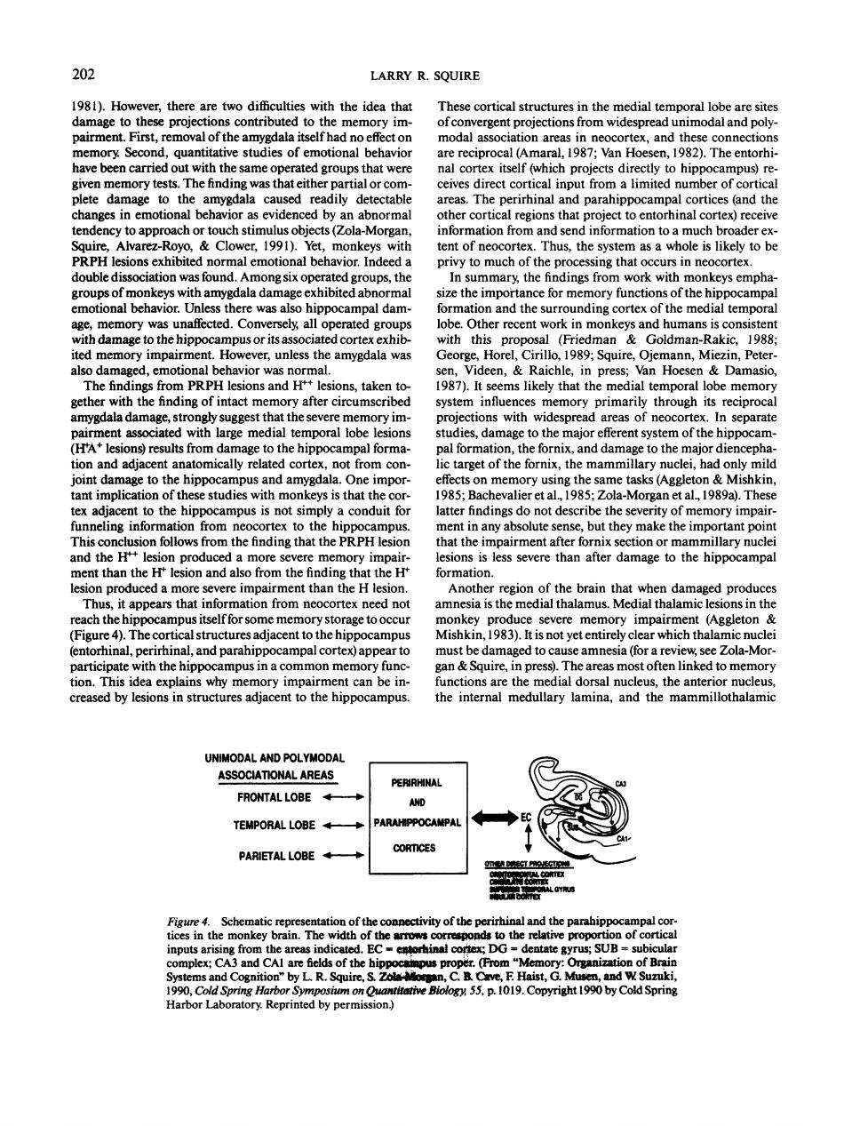
202 LARRY R.SQUIRE 1981).However,there are two difficulties with the idea that x and thm ory Seco stud ies of d 1987 Van Ho 1982).The entorhi ven meme ry tests The findin was that eithe artial or co ical inpu and th d b endency to approach or touch stimul sobjects(亿ola-Morga oad. e of th to b ouble dissociat keys empha motional beavior. so hipp mpal dam ely all operat d gro obe.Ot er recent -ite e ited memory impair ever,unless the amygdala was 1989:Squire,Ojemann,Mic n,Peter taken to cther with the ing of intact me ory after circumscrib system influe primarily through its reciproca iated with large medial temporal lobe to the major efferent system of the hi am s)re ults from da to the hip al form the fornix and damage to the m not ae to the hippo ampus a and amygdala.One imp on mem ory using 8 th same tasks (Aggleton&Mishkin nt to he hir s is not sim rfindings do not des icribe thes of me on from tex to .bu ute sense。. t they make the impc tant p the H++l is less severe than after damage to the hipp campal than hus. pears tha info 4)The es adia Mishkin.983)s inal,perirhin and parahipp ampal cortex)app maged to amnesia (for review sec Zola-Mor ion This idea explains why mer ment can he in functions are the medial do the creased by lesions in structures adjacent to the hippocampus. the internal medullary lamina,and the mammillothalami UNIMODAL AND POLYMODA ASSOCIATIONAL AREAS FRONTAL LOBE TEMPORAL LOBE PARIETAL LOBE ity ofthe perirhir d the the fro SUB CAl ar L R. 990.Cold Harbor Laboratory.Reprinted by permis ion) 202 LARRY R. SQUIRE 1981). However, there are two difficulties with the idea that damage to these projections contributed to the memory impairment. First, removal of the amygdala itself had no effect on memory. Second, quantitative studies of emotional behavior have been carried out with the same operated groups that were given memory tests. The finding was that either partial or complete damage to the amygdala caused readily detectable changes in emotional behavior as evidenced by an abnormal tendency to approach or touch stimulus objects (Zola-Morgan, Squire, Alvarez-Royo, & Clower, 1991). Yet, monkeys with PRPH lesions exhibited normal emotional behavior. Indeed a double dissociation was found. Among six operated groups, the groups of monkeys with amygdala damage exhibited abnormal emotional behavior. Unless there was also hippocampal damage, memory was unaffected. Conversely, all operated groups with damage to the hippocampus or its associated cortex exhibited memory impairment. However, unless the amygdala was also damaged, emotional behavior was normal. The findings from PRPH lesions and H++ lesions, taken together with the finding of intact memory after circumscribed amygdala damage, strongly suggest that the severe memory impairment associated with large medial temporal lobe lesions (tTA* lesions) results from damage to the hippocampal formation and adjacent anatomically related cortex, not from conjoint damage to the hippocampus and amygdala. One important implication of these studies with monkeys is that the cortex adjacent to the hippocampus is not simply a conduit for funneling information from neocortex to the hippocampus. This conclusion follows from the finding that the PRPH lesion and the H4 " 1 " lesion produced a more severe memory impairment than the IF lesion and also from the finding that the H+ lesion produced a more severe impairment than the H lesion. Thus, it appears that information from neocortex need not reach the hippocampus itself for some memory storage to occur (Figure 4). The cortical structures adjacent to the hippocampus (entorhinal, perirhinal, and parahippocampal cortex) appear to participate with the hippocampus in a common memory function. This idea explains why memory impairment can be increased by lesions in structures adjacent to the hippocampus. These cortical structures in the medial temporal lobe are sites of convergent projections from widespread unimodal and polymodal association areas in neocortex, and these connections are reciprocal (Amaral, 1987; Van Hoesen, 1982). The entorhinal cortex itself (which projects directly to hippocampus) receives direct cortical input from a limited number of cortical areas. The perirhinal and parahippocampal cortices (and the other cortical regions that project to entorhinal cortex) receive information from and send information to a much broader extent of neocortex. Thus, the system as a whole is likely to be privy to much of the processing that occurs in neocortex. In summary, the findings from work with monkeys emphasize the importance for memory functions of the hippocampal formation and the surrounding cortex of the medial temporal lobe. Other recent work in monkeys and humans is consistent with this proposal (Friedman & Goldman-Rakic, 1988; George, Horel, Cirillo, 1989; Squire, Ojemann, Miezin, Petersen, Videen, & Raichle, in press; Van Hoesen & Damasio, 1987). It seems likely that the medial temporal lobe memory system influences memory primarily through its reciprocal projections with widespread areas of neocortex. In separate studies, damage to the major efferent system of the hippocampal formation, the fornix, and damage to the major diencephalic target of the fornix, the mammillary nuclei, had only mild effects on memory using the same tasks (Aggleton & Mishkin, 1985; Bachevalier et al., 1985; Zola-Morgan et al., 1989a). These latter findings do not describe the severity of memory impairment in any absolute sense, but they make the important point that the impairment after fornix section or mammillary nuclei lesions is less severe than after damage to the hippocampal formation. Another region of the brain that when damaged produces amnesia is the medial thalamus. Medial thalamic lesions in the monkey produce severe memory impairment (Aggleton & Mishkin, 1983). It is not yet entirely clear which thalamic nuclei must be damaged to cause amnesia (for a review, see Zola-Morgan & Squire, in press). The areas most often linked to memory functions are the medial dorsal nucleus, the anterior nucleus, the internal medullary lamina, and the mammillothalamic UNIMODAL AND POLYMODAL ASSOCIATIONAL AREAS FRONTAL LOBE TEMPORAL LOBE PARIETAL LOBE Figure 4. Schematic representation of the connectivity of the perirhinal and the parahippocampal cortices in the monkey brain. The width of the arrows corresponds to the relative proportion of cortical inputs arising from the areas indicated. EC - eatorhinal cortex; DC = dentate gyrus; SUB = subicular complex; CA3 and CA1 are fields of the hippocanpus proper. (From "Memory: Organization of Brain Systems and Cognition" by L. R. Squire, & Zo1» Mocgin, C B. Cave, F. Haist, G. Musen, and W Suzuki, 1990, Cold Spring Harbor Symposium on Quantitative Biology, 55, p. 1019. Copyright 1990 by Cold Spring Harbor Laboratory. Reprinted by permission.)