正在加载图片...

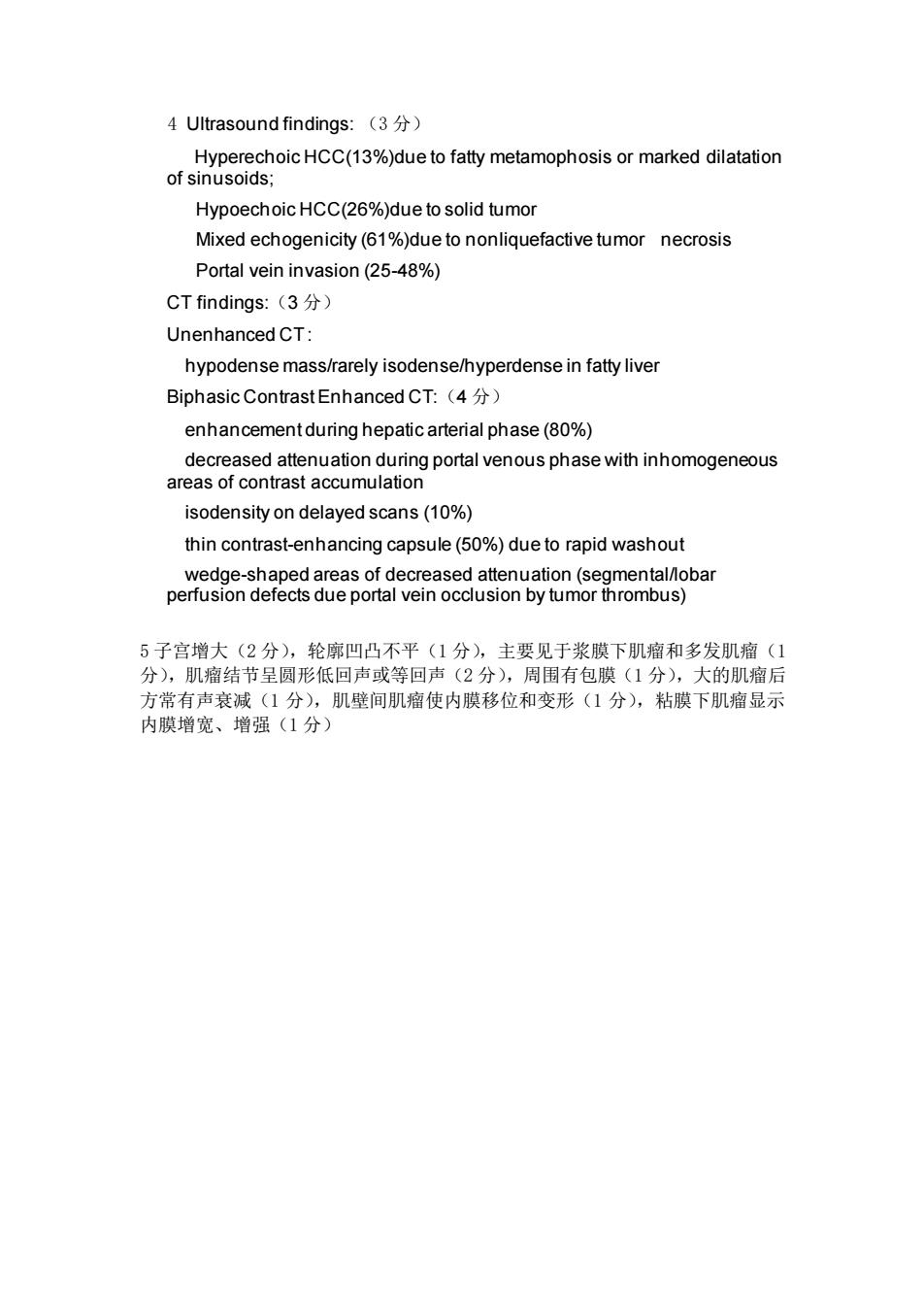
4 Ultrasound findings:(3 Hyperechoic HCC(13%)due to fatty metamophosis or marked dilatation of sinusoids; Hypoechoic HCC(26%)due to solid tumor Mixed echogenicity(61%)due to nonliquefactive tumor necrosis Portal vein invasion(25-48%) CT findings::(3分) Unenhanced CT: hypodense mass/rarely isodense/hyperdense in fatty liver Biphasic Contrast Enhanced CT:(4) enhancementduring hepatic arterial phase(80%) isodensity on delayed scans(10%) thin contrast-enhancing capsule(50%)due to rapid washout 5子宫增大(2分),轮廓凹凸不平(1分,主要见于浆膜下肌瘤和多发肌瘤( 分),肌瘤结节呈圆形低回声或等回声(2分),周围有包膜(1分),大的肌瘤后 方常有声衰减(1分),肌壁间肌瘤使内膜移位和变形(1分),粘膜下肌瘤显示 内膜增宽、增强(1分)4 Ultrasound findings: (3 分) Hyperechoic HCC(13%)due to fatty metamophosis or marked dilatation of sinusoids; Hypoechoic HCC(26%)due to solid tumor Mixed echogenicity (61%)due to nonliquefactive tumor necrosis Portal vein invasion (25-48%) CT findings:(3 分) Unenhanced CT : hypodense mass/rarely isodense/hyperdense in fatty liver Biphasic Contrast Enhanced CT:(4 分) enhancement during hepatic arterial phase (80%) decreased attenuation during portal venous phase with inhomogeneous areas of contrast accumulation isodensity on delayed scans (10%) thin contrast-enhancing capsule (50%) due to rapid washout wedge-shaped areas of decreased attenuation (segmental/lobar perfusion defects due portal vein occlusion by tumor thrombus) 5 子宫增大(2 分),轮廓凹凸不平(1 分),主要见于浆膜下肌瘤和多发肌瘤(1 分),肌瘤结节呈圆形低回声或等回声(2 分),周围有包膜(1 分),大的肌瘤后 方常有声衰减(1 分),肌壁间肌瘤使内膜移位和变形(1 分),粘膜下肌瘤显示 内膜增宽、增强(1 分)