正在加载图片...

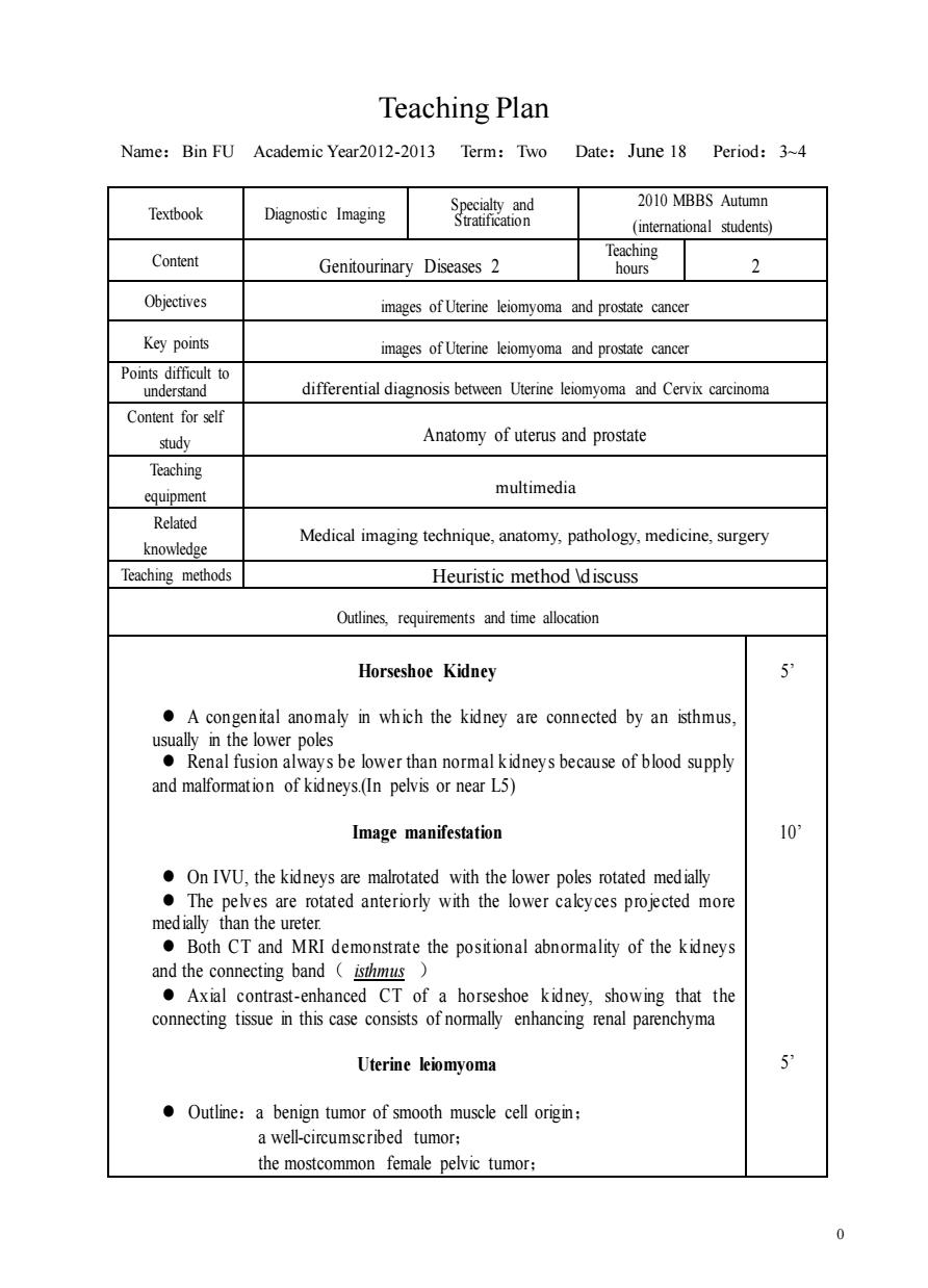
Teaching Plan Name:Bin FU Academic Year2012-2013 Term:Two Date:June 18 Period:3~4 Textbook Diagnostic Imaging 2010 MBBS Autumn (intemational students) Content Teaching Genitourinary Diseases 2 hours 2 Objectives images of Uterine leiomyoma and prostate cancer Key points images of Uterine leiomyoma and prostate cancer Points difficult to understand differential diagnosis between Uterine leiomyoma and Cervix carcinoma Content for self study Anatomy of uterus and prostate Teaching multimedia Related knowledge Medical imaging technique,anatomy,pathology,medicine,surgery Teaching methods Heuristic method discuss tinesrequrements and time alltion Horseshoe Kidney A congenital anomaly in which the kidney are connected by an isthmus usually in the oles ●Renal fusi of kidneys(In pelvis or near L5) Image manifestation 10 OnIVU,the kidneys are malrotated with the lower poles rotated medially The pelves are rotated anteriorly with the lower calcyces projected more medially than the ureter. Both CT and MRI demonstrate the positional abnormality of the kidneys and the connecting band isthmus ·Axial comrast-enhanced CT,showing that the connecting tissue in this case consists of normally enhancing renal parenchyma Uterine liomyoma Outline:a benign tumor of smooth muscle cell origin: a well-circumscribed tumor: the mostcommon female pelvic tumor: 00 Teaching Plan Name:Bin FU Academic Year2012-2013 Term:Two Date:June 18 Period:3~4 Textbook Diagnostic Imaging Specialty and Stratification 2010 MBBS Autumn (international students) Content Genitourinary Diseases 2 Teaching hours 2 Objectives images of Uterine leiomyoma and prostate cancer Key points images of Uterine leiomyoma and prostate cancer Points difficult to understand differential diagnosis between Uterine leiomyoma and Cervix carcinoma Content for self study Anatomy of uterus and prostate Teaching equipment multimedia Related knowledge Medical imaging technique, anatomy, pathology, medicine, surgery Teaching methods Heuristic method \discuss Outlines, requirements and time allocation Horseshoe Kidney ⚫ A congenital anomaly in wh ich the kidney are connected by an isthmus, usually in the lower poles ⚫ Renal fusion always be lower than normal kidneys because of blood supply and malformation of kidneys.(In pelvis or near L5) Image manifestation ⚫ On IVU, the kidneys are malrotated with the lower poles rotated medially ⚫ The pelves are rotated anteriorly with the lower calcyces projected more medially than the ureter. ⚫ Both CT and MRI demonstrate the positional abnormality of the kidneys and the connecting band( isthmus ) ⚫ Axial contrast-enhanced CT of a horseshoe kidney, showing that the connecting tissue in this case consists of normally enhancing renal parenchyma Uterine leiomyoma ⚫ Outline:a benign tumor of smooth muscle cell origin; a well-circumscribed tumor; the mostcommon female pelvic tumor; 5’ 10’ 5’