正在加载图片...
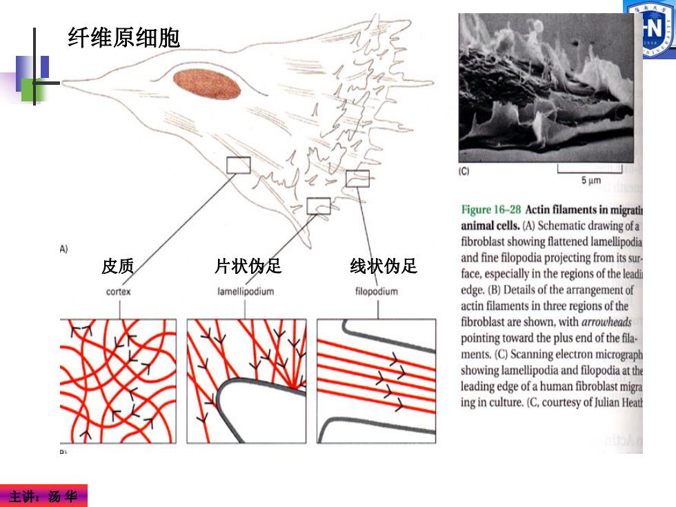
纤维原细胞 5μm Figure 16-28 Actin filaments in migrati animal cells.(A)Schematic drawing ofa fibroblast showing flattened lamellipodia 皮质 片状伪足 线状伪足 and fine filopodia projecting from its sur. face,especially in the regions of the leadi cortex lamellipodium filopodium edge.(B)Details of the arrangement of actin filaments in three regions of the fibroblast are shown,with arrowheads pointing toward the plus end of the fila. ments.(C)Scanning electron micrograph showing lamellipodia and filopodia at the leading edge of a human fibroblast migra ing in culture.(C.courtesy of Julian Heat 主讲汤华主讲:汤 华 皮质 片状伪足 线状伪足 纤维原细胞