正在加载图片...

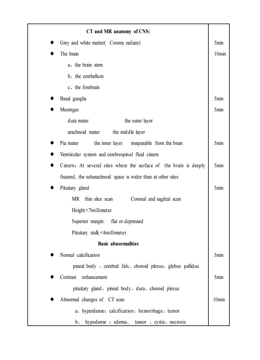
CT and MR anatomy of CNS: Grey and white matter(Corona radiate) 5min ·The brain 10min a、the brain stem b、the cerebellum c、the forebrain ●Basal ganglia 5min ·Meninges 5min dura mater the outer layer arachnoid mater the middle layer ●Pia mater the inner layer inseparable from the brain 5min Ventricular system and cerebrospinal fluid cistem Cistern:At several sites where the surface of the brain is deeply 5min fissured,the subarachnoid space is wider than at other sites. ●Pituitary gland 5min MR thin slice scan Coronal and sagittal scan Height:<7millimeter Superior margin:flat or depressed Pituitary stalk:<4millimeter Basic abnormalities ·Nomal calcification 5min pineal body、cerebral falx、choroid plexus、globus pallidus Contrast enhancement 5min pituitary gland、pineal body、dura、choroid plexus Abnormal changes of CT scan 10min a、hyperdense:calcification、hemorrhage、tumor b、hypodense:edema、tumor、cystis、necrosis CT and MR anatomy of CNS: ⚫ Grey and white matter( Corona radiate) ⚫ The brain a、the brain stem b、the cerebellum c、the forebrain ⚫ Basal ganglia ⚫ Meninges dura mater the outer layer arachnoid mater the mid dle layer ⚫ Pia mater the inner layer inseparable from the brain ⚫ Ventricular system and cerebrospinal fluid cistern ⚫ Cistern:At several sites where the surface of the brain is deeply fissured, the subarachnoid space is wider than at other sites. ⚫ Pituitary gland MR thin slice scan Coronal and sagittal scan Height:<7millimeter Superior margin: flat or depressed Pituitary stalk:<4millimeter Basic abnormalities ⚫ Normal calcification pineal body 、cerebral falx、choroid plexus、globus pallidus ⚫ Contrast enhancement pituitary gland、pineal body、dura、choroid plexus ⚫ Abnormal changes of CT scan a、hyperdense:calcification、hemorrhage、tumor b、 hypodense :edema、 tumor 、cystis、necrosis 5min 10min 5min 5min 5min 5min 5min 5min 5min 10min