正在加载图片...

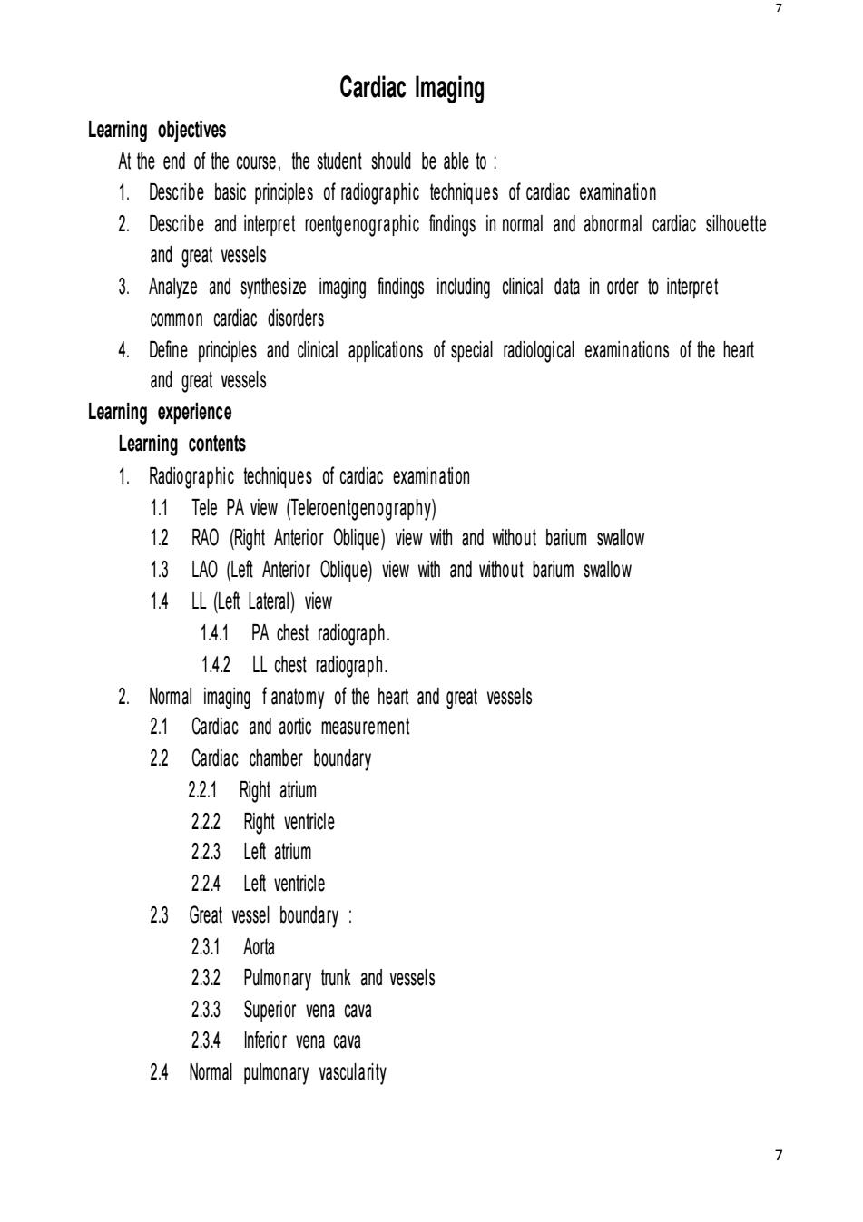
Cardiac Imaging Learing objectives At the end of the course,the student should be able to 1.Describe basic principles of radiographic techniqes of cardiac examination 2.Describe and interpret graphic findings in normal and abnormal cardiac sihouette and great vessels 3.Analyze and synthesize imaging findings including clinical data in order to interpret common cardiac disorders 4.Defie principles and cinical applicai of special radiogical examintions of the heart and great vessels Leaming experience Learning contents 1.Radiographic techniques of cardiac examinaion 1.1 Tele PA view (Teleroentgenography) 1.2 RAO (Right Anterior Oblique)view with and without barium swallow 1.3 LAO (Left Anterior Oblique)view with and without barium swallow 1.4 LL (Left Lateral)view 1.4.1 PA chest radiograph. 1.4.2 LL chest radiograph. 2.Nomal imaging fanatomy of the heart and great vessels 2.1 Cardiac and aortic measurement 2.2 Cardiac chamber boundary 2.2.1 Right atrium 2.2.2 Right ventricle 2.2.3 Left atrium 2.2.4 Left ventricle 2.3 Great vessel boundary 2.3.1 Aorta 2.3.2 Pulmonary trunk and vessels 2.3.3 Superior vena cava 2.3.4 Inferior vena cava 2.4 Normal pulmonary vascularity7 7 Cardiac Imaging Learning objectives At the end of the course, the student should be able to : 1. Describe basic principles of radiographic techniques of cardiac examination 2. Describe and interpret roentgenographic findings in normal and abnormal cardiac silhouette and great vessels 3. Analyze and synthesize imaging findings including clinical data in order to interpret common cardiac disorders 4. Define principles and clinical applications of special radiological examinations of the heart and great vessels Learning experience Learning contents 1. Radiographic techniques of cardiac examination 1.1 Tele PA view (Teleroentgenography) 1.2 RAO (Right Anterior Oblique) view with and without barium swallow 1.3 LAO (Left Anterior Oblique) view with and without barium swallow 1.4 LL (Left Lateral) view 1.4.1 PA chest radiograph. 1.4.2 LL chest radiograph. 2. Normal imaging f anatomy of the heart and great vessels 2.1 Cardiac and aortic measurement 2.2 Cardiac chamber boundary 2.2.1 Right atrium 2.2.2 Right ventricle 2.2.3 Left atrium 2.2.4 Left ventricle 2.3 Great vessel boundary : 2.3.1 Aorta 2.3.2 Pulmonary trunk and vessels 2.3.3 Superior vena cava 2.3.4 Inferior vena cava 2.4 Normal pulmonary vascularity