正在加载图片...
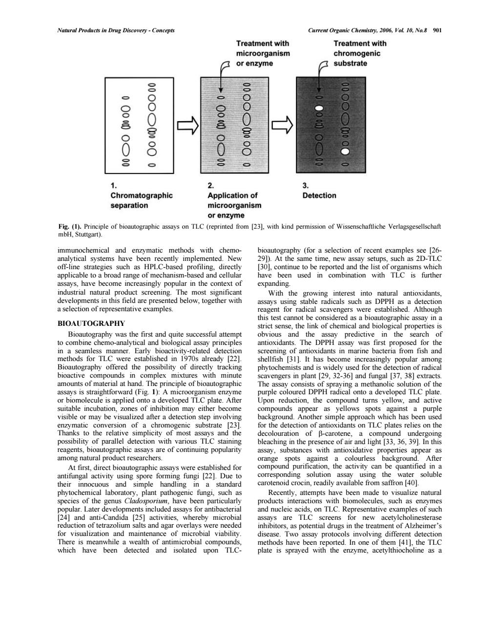
Natural Products in Drug Discovery-Concept Current Organic Chemistry.2006.VoL 10,No.8 901 Treatment with Treatment with rganism ogenic 7 or enzyme substrate S omatographic Application of Detection separation or enzyme inip of biooraphc on TIC (reprind fom with kind permission of Wisenschaftiche Verlagxgesellschaft 9 enzymatic hods with egies PLC- ing 30 popular in the context of expanding ning ith antioxidant rs we BIOAUTOGRAPHY the d fo n fish and the possibility of directly tra kin nd is ction of radic hand.Th cipe of bioauto tion of th A m ped TLC the om spots genic substrate 23 of parallel detection with ious TLC stai continuing popularity assay nces tographic the activity can carotenoid crocin.readily available from saffron 40 sucn as Later vel s for antibact on TLC.Represe tative c s of suck salts and agar ere ne ded inhibito re m ssay proto which have been detected and isolated upon TLONatural Products in Drug Discovery - Concepts Current Organic Chemistry, 2006, Vol. 10, No.8 901 immunochemical and enzymatic methods with chemoanalytical systems have been recently implemented. New off-line strategies such as HPLC-based profiling, directly applicable to a broad range of mechanism-based and cellular assays, have become increasingly popular in the context of industrial natural product screening. The most significant developments in this field are presented below, together with a selection of representative examples. BIOAUTOGRAPHY Bioautography was the first and quite successful attempt to combine chemo-analytical and biological assay principles in a seamless manner. Early bioactivity-related detection methods for TLC were established in 1970s already [22]. Bioautography offered the possibility of directly tracking bioactive compounds in complex mixtures with minute amounts of material at hand. The principle of bioautographic assays is straightforward (Fig. 1): A microorganism enzyme or biomolecule is applied onto a developed TLC plate. After suitable incubation, zones of inhibition may either become visible or may be visualized after a detection step involving enzymatic conversion of a chromogenic substrate [23]. Thanks to the relative simplicity of most assays and the possibility of parallel detection with various TLC staining reagents, bioautographic assays are of continuing popularity among natural product researchers. At first, direct bioautographic assays were established for antifungal activity using spore forming fungi [22]. Due to their innocuous and simple handling in a standard phytochemical laboratory, plant pathogenic fungi, such as species of the genus Cladosporium, have been particularly popular. Later developments included assays for antibacterial [24] and anti-Candida [25] activities, whereby microbial reduction of tetrazolium salts and agar overlays were needed for visualization and maintenance of microbial viability. There is meanwhile a wealth of antimicrobial compounds, which have been detected and isolated upon TLCbioautography (for a selection of recent examples see [26- 29]). At the same time, new assay setups, such as 2D-TLC [30], continue to be reported and the list of organisms which have been used in combination with TLC is further expanding. With the growing interest into natural antioxidants, assays using stable radicals such as DPPH as a detection reagent for radical scavengers were established. Although this test cannot be considered as a bioautographic assay in a strict sense, the link of chemical and biological properties is obvious and the assay predictive in the search of antioxidants. The DPPH assay was first proposed for the screening of antioxidants in marine bacteria from fish and shellfish [31]. It has become increasingly popular among phytochemists and is widely used for the detection of radical scavengers in plant [29, 32-36] and fungal [37, 38] extracts. The assay consists of spraying a methanolic solution of the purple coloured DPPH radical onto a developed TLC plate. Upon reduction, the compound turns yellow, and active compounds appear as yellows spots against a purple background. Another simple approach which has been used for the detection of antioxidants on TLC plates relies on the decolouration of b-carotene, a compound undergoing bleaching in the presence of air and light [33, 36, 39]. In this assay, substances with antioxidative properties appear as orange spots against a colourless background. After compound purification, the activity can be quantified in a corresponding solution assay using the water soluble carotenoid crocin, readily available from saffron [40]. Recently, attempts have been made to visualize natural products interactions with biomolecules, such as enzymes and nucleic acids, on TLC. Representative examples of such assays are TLC screens for new acetylcholinesterase inhibitors, as potential drugs in the treatment of Alzheimer’s disease. Two assay protocols involving different detection methods have been reported. In one of them [41], the TLC plate is sprayed with the enzyme, acetylthiocholine as a Fig. (1). Principle of bioautographic assays on TLC (reprinted from [23], with kind permission of Wissenschaftliche Verlagsgesellschaft mbH, Stuttgart)