正在加载图片...
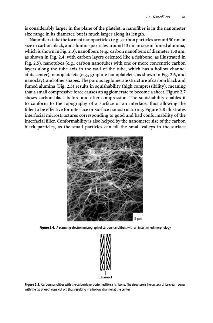
2.3 Nanofillers 41 is considerably larger in the plane of the platelet;a nanofiber is in the nanometer size range in its diameter,but is much larger along its length. Nanofillers take the form ofnanoparticles (e.g.,carbon particles around 30 nm in size in carbon black,and alumina particles around 13 nm in size in fumed alumina, which is shown in Fig.2.3),nanofibers(e.g.,carbon nanofibers of diameter 150nm, as shown in Fig.2.4,with carbon layers oriented like a fishbone,as illustrated in Fig.2.5),nanotubes (e.g.,carbon nanotubes with one or more concentric carbon layers along the tube axis in the wall of the tube,which has a hollow channel at its center),nanoplatelets(e.g.,graphite nanoplatelets,as shown in Fig.2.6,and nanoclay),and other shapes.The porous agglomerate structure ofcarbon black and fumed alumina(Fig.2.3)results in squishability (high compressibility),meaning that a small compressive force causes an agglomerate to become a sheet.Figure 2.7 shows carbon black before and after compression.The squishability enables it to conform to the topography of a surface or an interface,thus allowing the filler to be effective for interface or surface nanostructuring.Figure 2.8 illustrates interfacial microstructures corresponding to good and bad conformability of the interfacial filler.Conformability is also helped by the nanometer size of the carbon black particles,as the small particles can fill the small valleys in the surface 2m Figure 2.4.A scanning electron micrograph of carbon nanofibers with an intertwined morphology Channel Figure 2.5.Carbon nanofiber with the carbon layers oriented like a fishbone.The structure is like a stack of ice cream cones with the tip of each cone cut off,thus resulting in a hollow channel at the center2.3 Nanofillers 41 is considerably larger in the plane of the platelet; a nanofiber is in the nanometer size range in its diameter, but is much larger along its length. Nanofillerstaketheformofnanoparticles(e.g.,carbonparticlesaround30nmin size in carbon black, and alumina particles around 13nm in size in fumed alumina, which is shown in Fig. 2.3), nanofibers (e.g., carbon nanofibers of diameter 150nm, as shown in Fig. 2.4, with carbon layers oriented like a fishbone, as illustrated in Fig. 2.5), nanotubes (e.g., carbon nanotubes with one or more concentric carbon layers along the tube axis in the wall of the tube, which has a hollow channel at its center), nanoplatelets (e.g., graphite nanoplatelets, as shown in Fig. 2.6, and nanoclay),andothershapes.Theporousagglomeratestructureofcarbonblackand fumed alumina (Fig. 2.3) results in squishability (high compressibility), meaning that a small compressive force causes an agglomerate to become a sheet. Figure 2.7 shows carbon black before and after compression. The squishability enables it to conform to the topography of a surface or an interface, thus allowing the filler to be effective for interface or surface nanostructuring. Figure 2.8 illustrates interfacial microstructures corresponding to good and bad conformability of the interfacial filler. Conformability is also helped by the nanometer size of the carbon black particles, as the small particles can fill the small valleys in the surface Figure 2.4. A scanning electron micrograph of carbon nanofibers with an intertwined morphology Figure 2.5. Carbon nanofiber with the carbon layers oriented like a fishbone. The structure is like a stack of ice cream cones with the tip of each cone cut off, thus resulting in a hollow channel at the center