正在加载图片...
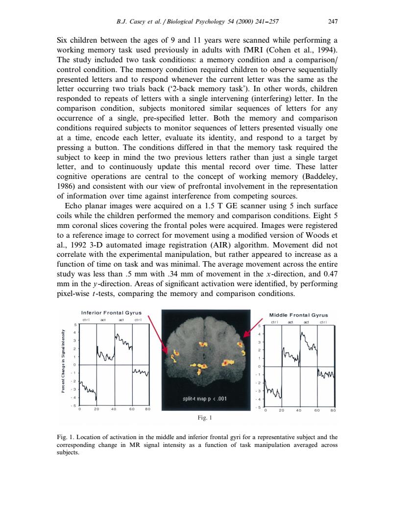
B.J.Casey et al.Biological Psychology 54(2000)241-257 Six children between the ages of 9 and 11 years were scanned while performing a working memory task used previously in adults with fMRI (Cohen et al.,1994). The study included two task conditions:a memory condition and a comparison/ control condition.The memory condition required children to observe sequentially as the 1uo9u7qN。 ever the current lette e same responded to repeats of letters with a single intervening (interfering)letter.In the comparison condition,subjects monitored similar sequences of letters for any occurrence of a single,pre-specified letter.Both the memory and comparison conditions required su cts to monitor s of letters presented visually on at a time,encode cach letter,evaluate sidentity,and respond to a target by pressing a button.The conditions differed in that the memory task required the subject to keep in mind the two previous letters rather than just a single target letter,and to continuously update this mental record over time.These latter operat cent al to the conce mory (Baddeley 6and consistent with our view of prefrontal involvement in the eprese of information over time against interference from competing sources. Echo planar images were acquired on a 1.5 T GE scanner using 5 inch surface coils while the children performed the memory and comparison conditions.Eight 5 mm coronal slices covering the frontal poles were acquired. Images were registered to a reference image to correct for movement using a modified version of Woods et al.,1992 3-D automated image registration (AIR)algorithm.Movement did not correlate with the experimental manipulation,but rather appeared to increase as a function of time on task and was minimal.The average mo ement across the entire study was less than .5 mm with.34 mm of mo ent in the x-dire ction,and 0.47 mm in the y-direction.Areas of significant activation were identified,by performing pixel-wise t-tests,comparing the memory and comparison conditions. Interior Frontal Gyru Middle Frontal Gyrus M Fig.I Fig.I.Location of activ in the middle and inferior f ntal gyri for an th subjects.B.J. Casey et al. / Biological Psychology 54 (2000) 241–257 247 Six children between the ages of 9 and 11 years were scanned while performing a working memory task used previously in adults with fMRI (Cohen et al., 1994). The study included two task conditions: a memory condition and a comparison/ control condition. The memory condition required children to observe sequentially presented letters and to respond whenever the current letter was the same as the letter occurring two trials back (‘2-back memory task’). In other words, children responded to repeats of letters with a single intervening (interfering) letter. In the comparison condition, subjects monitored similar sequences of letters for any occurrence of a single, pre-specified letter. Both the memory and comparison conditions required subjects to monitor sequences of letters presented visually one at a time, encode each letter, evaluate its identity, and respond to a target by pressing a button. The conditions differed in that the memory task required the subject to keep in mind the two previous letters rather than just a single target letter, and to continuously update this mental record over time. These latter cognitive operations are central to the concept of working memory (Baddeley, 1986) and consistent with our view of prefrontal involvement in the representation of information over time against interference from competing sources. Echo planar images were acquired on a 1.5 T GE scanner using 5 inch surface coils while the children performed the memory and comparison conditions. Eight 5 mm coronal slices covering the frontal poles were acquired. Images were registered to a reference image to correct for movement using a modified version of Woods et al., 1992 3-D automated image registration (AIR) algorithm. Movement did not correlate with the experimental manipulation, but rather appeared to increase as a function of time on task and was minimal. The average movement across the entire study was less than .5 mm with .34 mm of movement in the x-direction, and 0.47 mm in the y-direction. Areas of significant activation were identified, by performing pixel-wise t-tests, comparing the memory and comparison conditions. Fig. 1. Location of activation in the middle and inferior frontal gyri for a representative subject and the corresponding change in MR signal intensity as a function of task manipulation averaged across subjects