正在加载图片...
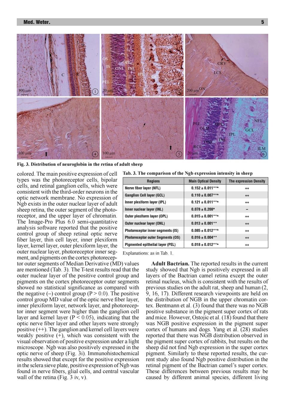
Med.Weter. 100m 100m 6 Fig.3.Distribution of neuroglobin in the retina of adult sheep colored.The main positive expression of cell Tab.3.The comparison of the Ngb expression intensity in sheep types was the photoreceptor cells,bipolar Regions Main Optical Density The expression Density cells,and retinal ganglion cells,which were Nerve fiber layer(NFL) 0.152±0.011*4 + consistent with the third-order neurons in the Ganglion Cell layer(GCL) 0.110±0.007*4 optic network membrane.No expression of Ngb exists in the outer nuclear layer of adult Inner plexiform layer(IPL) 0.121±0.011*4 sheep retina,the outer segment of the photo- Inner nuclear layer(INL) 0.078±0.2084 receptor,and the upper layer of chromatin. Outer plexiform layer(OPL) 0.015±0.001**4 + The Image-Pro Plus 6.0 semi-quantitative Outer nuclear layer(ONL) 0.013±0.001*+ + analysis software reported that the positive control group of sheep retinal optic nerve Photoreceptor inner segments(IS) 0.085±0.012*4 艹 fiber layer,thin cell layer,inner plexiform Photoreceptor outer Segments(OS) 0.016±0.004*+ layer,kernel layer,outer plexiform layer,the Pigmented epithelial layer(PEL) 0.018±0.012**4 + outer nuclear layer,photoreceptor inner seg- Explanations:as in Tab.1. ment,and pigments on the cortex photorecep- tor outer segments of Median Derivative(MD)values Adult Bactrian.The reported results in the current are mentioned(Tab.3).The T-test results read that the study showed that Ngb is positively expressed in all outer nuclear layer of the positive control group and layers of the Bactrian camel retina except the outer pigments on the cortex photoreceptor outer segments retinal nucleus,which is consistent with the results of showed no statistical significance as compared with previous studies on the adult rat,sheep and human(2, the negative(-)control group (P>0.0).The positive 9,16,17).Different research viewpoints are held on control group MD value of the optic nerve fiber layer, the distribution of NGB in the upper chromatin cor- inner plexiform layer,network layer,and photorecep- tex.Bentmann et al.(3)found that there was no NGB tor inner segment were higher than the ganglion cell positive substance in the pigment super cortex of rats layer and kernel layer(P<0.05),indicating that the and mice.However,Ostojic et al.(18)found that there optic nerve fiber layer and other layers were strongly was NGB positive expression in the pigment super positive(++).The ganglion and kernel cell layers were cortex of humans and dogs.Yang et al.(28)studies weakly positive (+)which was consistent with the reported that there was NGB distribution observed in visual observation of positive expression under a light the pigment super cortex of rabbits,but results on the microscope.Ngb was also positively expressed in the sheep did not find Ngb expression in the super cortex optic nerve of sheep(Fig.3i).Immunohistochemical pigment.Similarly to these reported results,the cur- results showed that except for the positive expression rent study also found Ngb positive distribution in the in the sclera sieve plate,positive expression of Ngb was retinal pigment of the Bactrian camel's super cortex. found in nerve fibers,glial cells,and central vascular These differences between previous results may be wall of the retina(Fig.3 iv,v). caused by different animal species,different livingMed. Weter. 5 colored. The main positive expression of cell types was the photoreceptor cells, bipolar cells, and retinal ganglion cells, which were consistent with the third-order neurons in the optic network membrane. No expression of Ngb exists in the outer nuclear layer of adult sheep retina, the outer segment of the photoreceptor, and the upper layer of chromatin. The Image-Pro Plus 6.0 semi-quantitative analysis software reported that the positive control group of sheep retinal optic nerve fiber layer, thin cell layer, inner plexiform layer, kernel layer, outer plexiform layer, the outer nuclear layer, photoreceptor inner segment, and pigments on the cortex photoreceptor outer segments of Median Derivative (MD) values are mentioned (Tab. 3). The T-test results read that the outer nuclear layer of the positive control group and pigments on the cortex photoreceptor outer segments showed no statistical significance as compared with the negative (–) control group (P > 0.0). The positive control group MD value of the optic nerve fiber layer, inner plexiform layer, network layer, and photoreceptor inner segment were higher than the ganglion cell layer and kernel layer (P < 0.05), indicating that the optic nerve fiber layer and other layers were strongly positive (++). The ganglion and kernel cell layers were weakly positive (+), which was consistent with the visual observation of positive expression under a light microscope. Ngb was also positively expressed in the optic nerve of sheep (Fig. 3i). Immunohistochemical results showed that except for the positive expression in the sclera sieve plate, positive expression of Ngb was found in nerve fibers, glial cells, and central vascular wall of the retina (Fig. 3 iv, v). Adult Bactrian. The reported results in the current study showed that Ngb is positively expressed in all layers of the Bactrian camel retina except the outer retinal nucleus, which is consistent with the results of previous studies on the adult rat, sheep and human (2, 9, 16, 17). Different research viewpoints are held on the distribution of NGB in the upper chromatin cortex. Bentmann et al. (3) found that there was no NGB positive substance in the pigment super cortex of rats and mice. However, Ostojic et al. (18) found that there was NGB positive expression in the pigment super cortex of humans and dogs. Yang et al. (28) studies reported that there was NGB distribution observed in the pigment super cortex of rabbits, but results on the sheep did not find Ngb expression in the super cortex pigment. Similarly to these reported results, the current study also found Ngb positive distribution in the retinal pigment of the Bactrian camel’s super cortex. These differences between previous results may be caused by different animal species, different living Tab. 3. The comparison of the Ngb expression intensity in sheep Regions Main Optical Density The expression Density Nerve fiber layer (NFL) 0.152 ± 0.011**▲ ++ Ganglion Cell layer (GCL) 0.110 ± 0.007**▲ ++ Inner plexiform layer (IPL) 0.121 ± 0.011**▲ ++ Inner nuclear layer (INL) 0.078 ± 0.208▲ – Outer plexiform layer (OPL) 0.015 ± 0.001**▲ ++ Outer nuclear layer (ONL) 0.013 ± 0.001** ++ Photoreceptor inner segments (IS) 0.085 ± 0.012**▲ ++ Photoreceptor outer Segments (OS) 0.016 ± 0.004** ++ Pigmented epithelial layer (PEL) 0.018 ± 0.012**▲ ++ Explanations: as in Tab. 1. Fig. 3. Distribution of neuroglobin in the retina of adult sheep