正在加载图片...
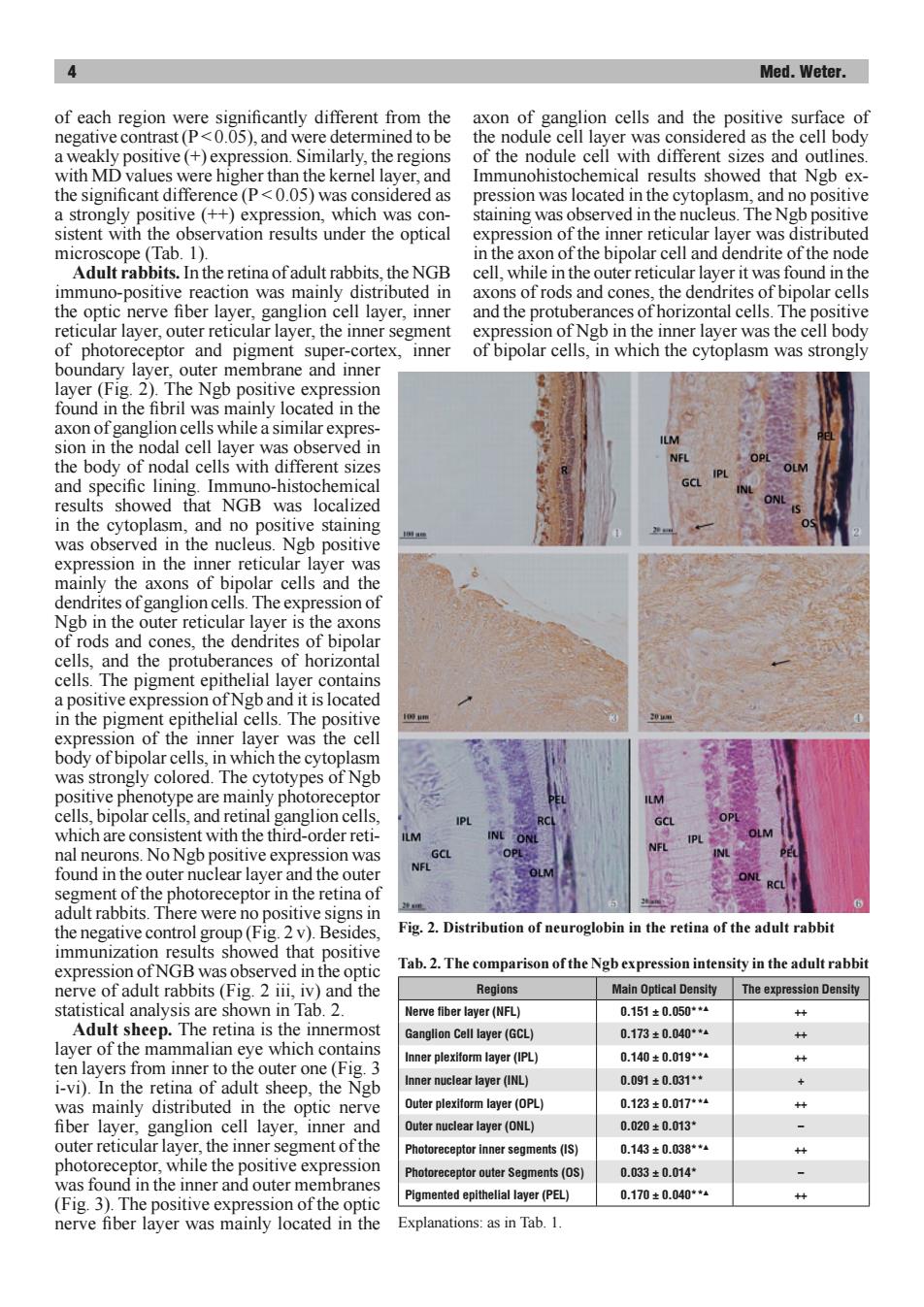
Med.Weter. of each region were significantly different from the axon of ganglion cells and the positive surface of negative contrast(P<0.05),and were determined to be the nodule cell layer was considered as the cell body a weakly positive (+)expression.Similarly,the regions of the nodule cell with different sizes and outlines. with MD values were higher than the kernel layer,and Immunohistochemical results showed that Ngb ex- the significant difference(P<0.05)was considered as pression was located in the cytoplasm,and no positive a strongly positive (++)expression,which was con- staining was observed in the nucleus.The Ngb positive sistent with the observation results under the optical expression of the inner reticular layer was distributed microscope (Tab.1). in the axon of the bipolar cell and dendrite of the node Adult rabbits.In the retina of adult rabbits,the NGB cell,while in the outer reticular layer it was found in the immuno-positive reaction was mainly distributed in axons of rods and cones,the dendrites of bipolar cells the optic nerve fiber layer,ganglion cell layer,inner and the protuberances of horizontal cells.The positive reticular layer,outer reticular layer,the inner segment expression of Ngb in the inner layer was the cell body of photoreceptor and pigment super-cortex,inner of bipolar cells,in which the cytoplasm was strongly boundary layer,outer membrane and inner layer(Fig.2).The Ngb positive expression found in the fibril was mainly located in the axon of ganglion cells while a similar expres- sion in the nodal cell layer was observed in the body of nodal cells with different sizes and specific lining.Immuno-histochemical results showed that NGB was localized in the cytoplasm,and no positive staining was observed in the nucleus.Ngb positive expression in the inner reticular layer was mainly the axons of bipolar cells and the dendrites of ganglion cells.The expression of Ngb in the outer reticular layer is the axons of rods and cones,the dendrites of bipolar cells,and the protuberances of horizontal cells.The pigment epithelial layer contains a positive expression of Ngb and it is located in the pigment epithelial cells.The positive expression of the inner layer was the cell body of bipolar cells,in which the cytoplasm was strongly colored.The cytotypes of Ngb positive phenotype are mainly photoreceptor cells,bipolar cells,and retinal ganglion cells, which are consistent with the third-order reti- nal neurons.No Ngb positive expression was found in the outer nuclear layer and the outer segment of the photoreceptor in the retina of adult rabbits.There were no positive signs in the negative control group(Fig.2 v).Besides. Fig.2.Distribution of neuroglobin in the retina of the adult rabbit immunization results showed that positive expression of NGB was observed in the optic Tab.2.The comparison of the Ngb expression intensity in the adult rabbit nerve of adult rabbits (Fig.2 iii,iv)and the Regions Main Optical Density The expression Density statistical analysis are shown in Tab.2. Nerve fiber layer(NFL) 0.151±0.050**4 x Adult sheep.The retina is the innermost Ganglion Cell layer(GCL) 0.173±0.040**4 x layer of the mammalian eye which contains Inner plexiform layer(IPL) 0.140±0.019*4 ten layers from inner to the outer one(Fig.3 米x i-vi).In the retina of adult sheep,the Ngb Inner nuclear layer(INL) 0.091±0.031◆+ + was mainly distributed in the optic nerve Outer plexiform layer(OPL) 0.123±0.017**“ x fiber layer,ganglion cell layer,inner and Outer nuclear layer(ONL) 0.020±0.013* outer reticular layer,the inner segment ofthe Photoreceptor inner segments(IS) 0.143±0.038**4 photoreceptor,while the positive expression Photoreceptor outer Segments(OS) 0.033±0.014 was found in the inner and outer membranes Pigmented epithelial layer(PEL) 0.170±0.040**1 (Fig.3).The positive expression of the optic nerve fiber layer was mainly located in the Explanations:as in Tab.1.4 Med. Weter. of each region were significantly different from the negative contrast (P < 0.05), and were determined to be a weakly positive (+) expression. Similarly, the regions with MD values were higher than the kernel layer, and the significant difference (P < 0.05) was considered as a strongly positive (++) expression, which was consistent with the observation results under the optical microscope (Tab. 1). Adult rabbits. In the retina of adult rabbits, the NGB immuno-positive reaction was mainly distributed in the optic nerve fiber layer, ganglion cell layer, inner reticular layer, outer reticular layer, the inner segment of photoreceptor and pigment super-cortex, inner boundary layer, outer membrane and inner layer (Fig. 2). The Ngb positive expression found in the fibril was mainly located in the axon of ganglion cells while a similar expression in the nodal cell layer was observed in the body of nodal cells with different sizes and specific lining. Immuno-histochemical results showed that NGB was localized in the cytoplasm, and no positive staining was observed in the nucleus. Ngb positive expression in the inner reticular layer was mainly the axons of bipolar cells and the dendrites of ganglion cells. The expression of Ngb in the outer reticular layer is the axons of rods and cones, the dendrites of bipolar cells, and the protuberances of horizontal cells. The pigment epithelial layer contains a positive expression of Ngb and it is located in the pigment epithelial cells. The positive expression of the inner layer was the cell body of bipolar cells, in which the cytoplasm was strongly colored. The cytotypes of Ngb positive phenotype are mainly photoreceptor cells, bipolar cells, and retinal ganglion cells, which are consistent with the third-order retinal neurons. No Ngb positive expression was found in the outer nuclear layer and the outer segment of the photoreceptor in the retina of adult rabbits. There were no positive signs in the negative control group (Fig. 2 v). Besides, immunization results showed that positive expression of NGB was observed in the optic nerve of adult rabbits (Fig. 2 iii, iv) and the statistical analysis are shown in Tab. 2. Adult sheep. The retina is the innermost layer of the mammalian eye which contains ten layers from inner to the outer one (Fig. 3 i-vi). In the retina of adult sheep, the Ngb was mainly distributed in the optic nerve fiber layer, ganglion cell layer, inner and outer reticular layer, the inner segment of the photoreceptor, while the positive expression was found in the inner and outer membranes (Fig. 3). The positive expression of the optic nerve fiber layer was mainly located in the axon of ganglion cells and the positive surface of the nodule cell layer was considered as the cell body of the nodule cell with different sizes and outlines. Immunohistochemical results showed that Ngb expression was located in the cytoplasm, and no positive staining was observed in the nucleus. The Ngb positive expression of the inner reticular layer was distributed in the axon of the bipolar cell and dendrite of the node cell, while in the outer reticular layer it was found in the axons of rods and cones, the dendrites of bipolar cells and the protuberances of horizontal cells. The positive expression of Ngb in the inner layer was the cell body of bipolar cells, in which the cytoplasm was strongly Tab. 2. The comparison of the Ngb expression intensity in the adult rabbit Regions Main Optical Density The expression Density Nerve fiber layer (NFL) 0.151 ± 0.050**▲ ++ Ganglion Cell layer (GCL) 0.173 ± 0.040**▲ ++ Inner plexiform layer (IPL) 0.140 ± 0.019**▲ ++ Inner nuclear layer (INL) 0.091 ± 0.031** + Outer plexiform layer (OPL) 0.123 ± 0.017**▲ ++ Outer nuclear layer (ONL) 0.020 ± 0.013* – Photoreceptor inner segments (IS) 0.143 ± 0.038**▲ ++ Photoreceptor outer Segments (OS) 0.033 ± 0.014* – Pigmented epithelial layer (PEL) 0.170 ± 0.040**▲ ++ Explanations: as in Tab. 1. Fig. 2. Distribution of neuroglobin in the retina of the adult rabbit