正在加载图片...
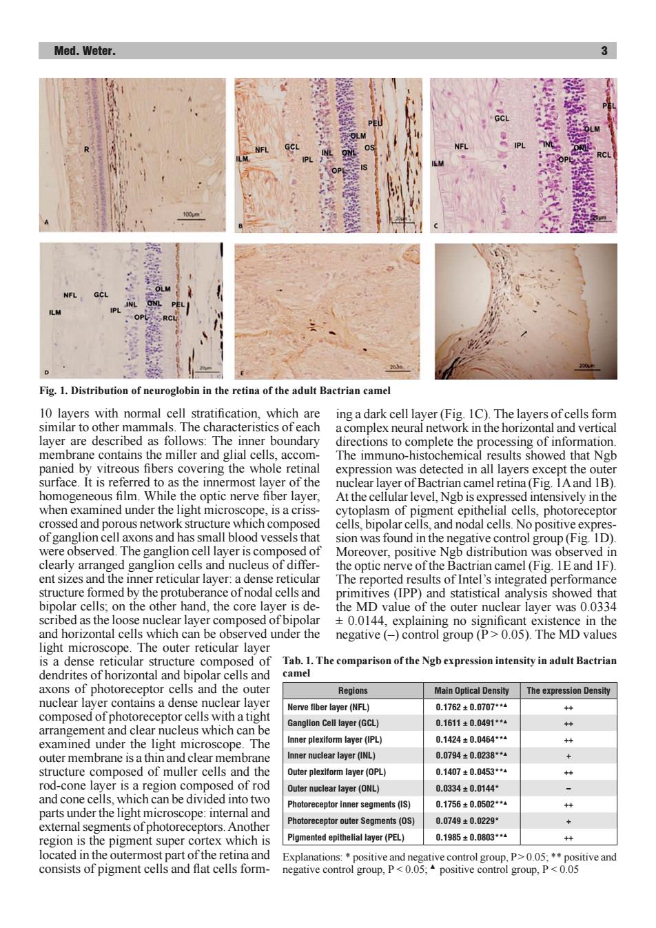
Med.Weter. Fig.1.Distribution of neuroglobin in the retina of the adult Bactrian camel 10 layers with normal cell stratification,which are ing a dark cell layer(Fig.1C).The layers of cells form similar to other mammals.The characteristics of each a complex neural network in the horizontal and vertical layer are described as follows:The inner boundary directions to complete the processing of information membrane contains the miller and glial cells,accom- The immuno-histochemical results showed that Ngb panied by vitreous fibers covering the whole retinal expression was detected in all layers except the outer surface.It is referred to as the innermost layer of the nuclear layer of Bactrian camel retina(Fig.1A and 1B). homogeneous film.While the optic nerve fiber layer, At the cellular level,Ngb is expressed intensively in the when examined under the light microscope,is a criss- cytoplasm of pigment epithelial cells,photoreceptor crossed and porous network structure which composed cells,bipolar cells,and nodal cells.No positive expres- of ganglion cell axons and has small blood vessels that sion was found in the negative control group (Fig.1D) were observed.The ganglion cell layer is composed of Moreover,positive Ngb distribution was observed in clearly arranged ganglion cells and nucleus of differ- the optic nerve of the Bactrian camel(Fig.1E and 1F). ent sizes and the inner reticular layer:a dense reticular The reported results of Intel's integrated performance structure formed by the protuberance of nodal cells and primitives (IPP)and statistical analysis showed that bipolar cells;on the other hand,the core layer is de- the MD value of the outer nuclear layer was 0.0334 scribed as the loose nuclear layer composed of bipolar +0.0144,explaining no significant existence in the and horizontal cells which can be observed under the negative (-)control group(P>0.05).The MD values light microscope.The outer reticular layer is a dense reticular structure composed of Tab.1.The comparison of the Ngb expression intensity in adult Bactrian dendrites of horizontal and bipolar cells and camel axons of photoreceptor cells and the outer Regions Main Optical Density The expression Density nuclear layer contains a dense nuclear layer Nerve fiber layer(NFL) 0.1762±0.0707*4 ++ composed of photoreceptor cells with a tight Ganglion Cell layer(GCL) 0.1611±0.0491**a ++ arrangement and clear nucleus which can be examined under the light microscope.The Inner plexiform layer(IPL) 0.1424±0.0464** outer membrane is a thin and clear membrane Inner nuclear layer(INL) 0.0794±0.0238**4 + structure composed of muller cells and the Outer plexiform layer(OPL) 0.1407±0.0453**4 ++ rod-cone layer is a region composed of rod Outer nuclear layer(ONL) 0.0334±0.0144 and cone cells,which can be divided into two Photoreceptor inner segments(IS) 0.1756±0.0502**4 ++ parts under the light microscope:internal and Photoreceptor outer Segments(OS) 0.0749±0.0229 external segments of photoreceptors.Another region is the pigment super cortex which is Pigmented epithelial layer(PEL) 0.1985±0.0803*4 ++ located in the outermost part of the retina and Explanations:*positive and negative control group,P>0.05;**positive and consists of pigment cells and flat cells form- negative control group,P<0.05;positive control group,P<0.05Med. Weter. 3 10 layers with normal cell stratification, which are similar to other mammals. The characteristics of each layer are described as follows: The inner boundary membrane contains the miller and glial cells, accompanied by vitreous fibers covering the whole retinal surface. It is referred to as the innermost layer of the homogeneous film. While the optic nerve fiber layer, when examined under the light microscope, is a crisscrossed and porous network structure which composed of ganglion cell axons and has small blood vessels that were observed. The ganglion cell layer is composed of clearly arranged ganglion cells and nucleus of different sizes and the inner reticular layer: a dense reticular structure formed by the protuberance of nodal cells and bipolar cells; on the other hand, the core layer is described as the loose nuclear layer composed of bipolar and horizontal cells which can be observed under the light microscope. The outer reticular layer is a dense reticular structure composed of dendrites of horizontal and bipolar cells and axons of photoreceptor cells and the outer nuclear layer contains a dense nuclear layer composed of photoreceptor cells with a tight arrangement and clear nucleus which can be examined under the light microscope. The outer membrane is a thin and clear membrane structure composed of muller cells and the rod-cone layer is a region composed of rod and cone cells, which can be divided into two parts under the light microscope: internal and external segments of photoreceptors. Another region is the pigment super cortex which is located in the outermost part of the retina and consists of pigment cells and flat cells forming a dark cell layer (Fig. 1C). The layers of cells form a complex neural network in the horizontal and vertical directions to complete the processing of information. The immuno-histochemical results showed that Ngb expression was detected in all layers except the outer nuclear layer of Bactrian camel retina (Fig. 1A and 1B). At the cellular level, Ngb is expressed intensively in the cytoplasm of pigment epithelial cells, photoreceptor cells, bipolar cells, and nodal cells. No positive expression was found in the negative control group (Fig. 1D). Moreover, positive Ngb distribution was observed in the optic nerve of the Bactrian camel (Fig. 1E and 1F). The reported results of Intel’s integrated performance primitives (IPP) and statistical analysis showed that the MD value of the outer nuclear layer was 0.0334 ± 0.0144, explaining no significant existence in the negative (–) control group (P > 0.05). The MD values Tab. 1. The comparison of the Ngb expression intensity in adult Bactrian camel Regions Main Optical Density The expression Density Nerve fiber layer (NFL) 0.1762 ± 0.0707**▲ ++ Ganglion Cell layer (GCL) 0.1611 ± 0.0491**▲ ++ Inner plexiform layer (IPL) 0.1424 ± 0.0464**▲ ++ Inner nuclear layer (INL) 0.0794 ± 0.0238**▲ + Outer plexiform layer (OPL) 0.1407 ± 0.0453**▲ ++ Outer nuclear layer (ONL) 0.0334 ± 0.0144* – Photoreceptor inner segments (IS) 0.1756 ± 0.0502**▲ ++ Photoreceptor outer Segments (OS) 0.0749 ± 0.0229* + Pigmented epithelial layer (PEL) 0.1985 ± 0.0803**▲ ++ Explanations: * positive and negative control group, P > 0.05; ** positive and negative control group, P < 0.05; ▲ positive control group, P < 0.05 Fig. 1. Distribution of neuroglobin in the retina of the adult Bactrian camel