正在加载图片...
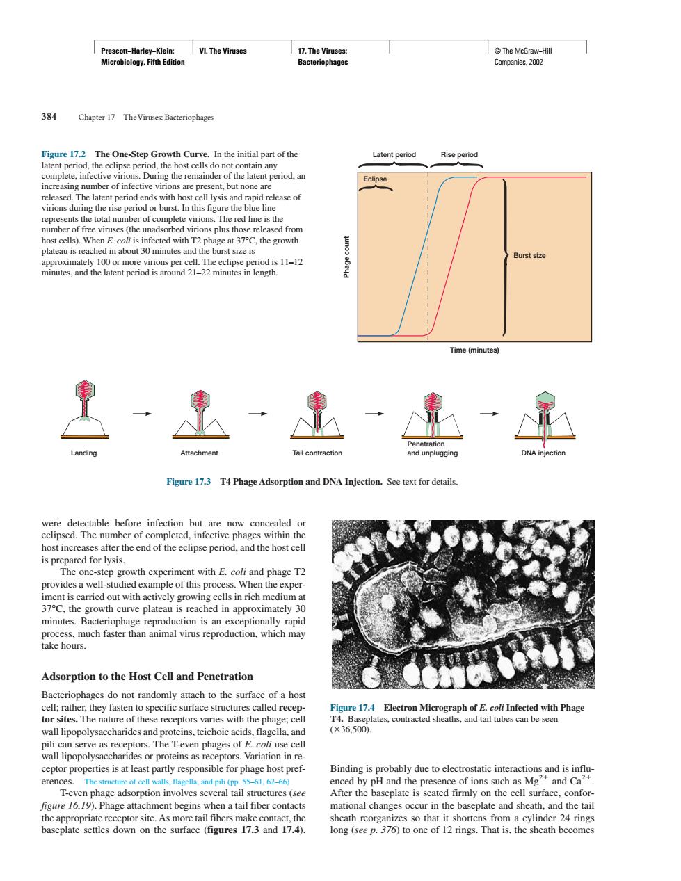
Chapter 17 The Viruses:Bacteriophages re 17 The One-Step G Laten period Rise period During the re atent p od.ar (the 100 11-12 Time(minutes 里速速业平 Figure 17.T4 Phage Adsorption and DNA Injection. is prepared for lysis. facrhamalisc Adsorption to the Host Cell and Penetration teriophages do not randomly attach to the surface of a host at时n ceptor properties is at least parly responsible for phage host pref- .19).Phagc atta ment begins when a tail fiber n mationl changes i the baseplate and sheath.and the tai long (ep7)to on of 12 rings.That is,the sheath becomePrescott−Harley−Klein: Microbiology, Fifth Edition VI. The Viruses 17. The Viruses: Bacteriophages © The McGraw−Hill Companies, 2002 Binding is probably due to electrostatic interactions and is influenced by pH and the presence of ions such as Mg2 and Ca2. After the baseplate is seated firmly on the cell surface, conformational changes occur in the baseplate and sheath, and the tail sheath reorganizes so that it shortens from a cylinder 24 rings long (see p. 376) to one of 12 rings. That is, the sheath becomes 384 Chapter 17 The Viruses: Bacteriophages were detectable before infection but are now concealed or eclipsed. The number of completed, infective phages within the host increases after the end of the eclipse period, and the host cell is prepared for lysis. The one-step growth experiment with E. coli and phage T2 provides a well-studied example of this process. When the experiment is carried out with actively growing cells in rich medium at 37°C, the growth curve plateau is reached in approximately 30 minutes. Bacteriophage reproduction is an exceptionally rapid process, much faster than animal virus reproduction, which may take hours. Adsorption to the Host Cell and Penetration Bacteriophages do not randomly attach to the surface of a host cell; rather, they fasten to specific surface structures called receptor sites. The nature of these receptors varies with the phage; cell wall lipopolysaccharides and proteins, teichoic acids, flagella, and pili can serve as receptors. The T-even phages of E. coli use cell wall lipopolysaccharides or proteins as receptors. Variation in receptor properties is at least partly responsible for phage host preferences. The structure of cell walls, flagella, and pili (pp. 55–61, 62–66) T-even phage adsorption involves several tail structures (see figure 16.19). Phage attachment begins when a tail fiber contacts the appropriate receptor site. As more tail fibers make contact, the baseplate settles down on the surface (figures 17.3 and 17.4). Time (minutes) Phage count Burst size Latent period Rise period Eclipse Figure 17.2 The One-Step Growth Curve. In the initial part of the latent period, the eclipse period, the host cells do not contain any complete, infective virions. During the remainder of the latent period, an increasing number of infective virions are present, but none are released. The latent period ends with host cell lysis and rapid release of virions during the rise period or burst. In this figure the blue line represents the total number of complete virions. The red line is the number of free viruses (the unadsorbed virions plus those released from host cells). When E. coli is infected with T2 phage at 37°C, the growth plateau is reached in about 30 minutes and the burst size is approximately 100 or more virions per cell. The eclipse period is 11–12 minutes, and the latent period is around 21–22 minutes in length. Landing Attachment Tail contraction Penetration and unplugging DNA injection Figure 17.3 T4 Phage Adsorption and DNA Injection. See text for details. Figure 17.4 Electron Micrograph of E. coli Infected with Phage T4. Baseplates, contracted sheaths, and tail tubes can be seen (36,500).��