正在加载图片...
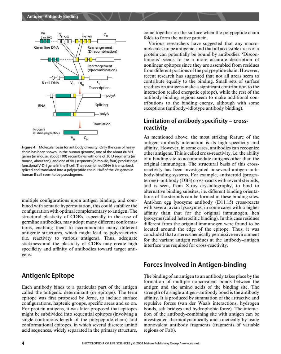
Anigen-Antibody Binding come together on the surface when the polypeptide chain 棉00g00-0900日 folds to form the native protein. Germ line DNA 0.出00- tinuous seems to be a more accurate description of nonlinear epitopes since they are assembled from residues on othe polypeptide chain.o has suggested that not all areas seem to V Dl.. residues on antigens make a significant contribution to the poyA al con Splicing aeeEno-oybag with some ☐-polyA Limitation of antibody specificity-cross reactivity As mentioned above,the most striking feature of the Figure4 Molecular basis for antibody diversity.Only the other antigens This is called cross-reactivity.Le.the ability of a binding site to accommodate antigens other than the ctpolypeptide chanHa munogen.The structural basis of this cross- one)-antibody (DB3)cross-reacts with sev and is seen,from X-ray crystallography.to bind to the stero multiple configurations upon antigen binding.and com D1115 with a higher iall in the affinity than that for the original immunogen,hen germline antibodies,may adopt many different conforma ysozyme(called heteroclitic binding).In this case residues eorigina un Th stickiness and the plasticity of CDRs may create high for the variant antigen residues at the antibody-antigen specificity and affinity of antibodies toward target anti interface was required for cross-reactivity. gens. Forces Involved in Antigen-binding Antigenic Epitope The binding ofanantigen to an antibody takes place by the tbonds between the e was first pi e to include surface ffinity It is produced by summation of the attractive and repulsive forces (van der Waals interactions,hydrogen mig ng e antibody-com ning site w th an ugen can b 、in which several discrete a acid sequences,widely separated in the primary structure. regions or Fab). ENCYCLOPEDIA OF LIFE SCENCES/2001Na shing Group /www.els.nemultiple configurations upon antigen binding, and combined with somatic hypermutation, this could stabilize the configuration with optimal complementary to antigen. The structural plasticity of CDRs, especially in the case of germline antibodies, may adopt many different conformations, enabling them to accommodate many different antigenic structures, which might lead to polyreactivity (i.e. reactivity to various antigens). Thus, adequate stickiness and the plasticity of CDRs may create high specificity and affinity of antibodies toward target antigens. Antigenic Epitope Each antibody binds to a particular part of the antigen called the antigenic determinant (or epitope). The term epitope was first proposed by Jerne, to include surface configurations, haptenic groups, specific areas and so on. For protein antigens, it was later proposed that epitopes might be subdivided into sequential epitopes (involving a single continuous length of the polypeptide chain) and conformational epitopes, in which several discrete amino acid sequences, widely separated in the primary structure, come together on the surface when the polypeptide chain folds to form the native protein. Various researchers have suggested that any macromolecule can be antigenic, and that all accessible areas of a protein can potentially be bound by antibodies. ‘Discontinuous’ seems to be a more accurate description of nonlinear epitopes since they are assembled from residues from different portions of the polypeptide chain. However, recent research has suggested that not all areas seem to contribute equally to the binding. Small sets of surface residues on antigens make a significant contribution to the interaction (called energetic epitope), while the rest of the antibody-binding regions seem to make additional contributions to the binding energy, although with some exceptions (antibody–idiotype antibody binding). Limitation of antibody specificity – crossreactivity As mentioned above, the most striking feature of the antigen–antibody interaction is its high specificity and affinity. However, in some cases, antibodies can recognize other antigens. This is called cross-reactivity, i.e. the ability of a binding site to accommodate antigens other than the original immunogen. The structural basis of this crossreactivity has been investigated in several antigen–antibody-binding systems. For example, antisteroid (progesterone)–antibody (DB3) cross-reacts with several steroids, and is seen, from X-ray crystallography, to bind to alternative binding subsites, i.e. different binding orientations of the steroids can be formed in these binding sites. Anti-hen egg lysozyme antibody (D11.15) cross-reacts with several avian lysozymes, in some cases with a higher affinity than that for the original immunogen, hen lysozyme (called heteroclitic binding). In this case residues different from the original immunogen were found to be located around the edge of the epitope. Thus, it was concluded that a stereochemically permissive environment for the variant antigen residues at the antibody–antigen interface was required for cross-reactivity. Forces Involved in Antigen-binding The binding of an antigen to an antibody takes place by the formation of multiple noncovalent bonds between the antigen and the amino acids of the binding site. The strength of a single antigen–antibody bond is the antibody affinity. It is produced by summation of the attractive and repulsive forces (van der Waals interactions, hydrogen bonds, salt bridges and hydrophobic force). The interaction of the antibody-combining site with antigen can be investigated thermodynamically and kinetically by using monovalent antibody fragments (fragments of variable regions or Fab). VH ( ˜ca 200) D(1˜20) J H(1˜6) CH Rearrangement (DJrecombination) Rearrangement (VJrecombination) CH Transcription DJH polyA Germ line DNA B cell DNA VH Splicing polyA Translation RNA Protein (H chain polypeptide) VH CH Figure 4 Molecular basis for antibody diversity. Only the case of heavy chain has been shown. In the human genome, one of the about 80 VH genes (in mouse, about 100) recombines with one of 30 D segments (in mouse, about ten), and one of six J segments (in mouse, four) producing a functional V-D-J gene in the B cell. The recombined DNA is transcribed, spliced and translated into a polypeptide chain. Half of the VH genes in human B cell seem to be pseudogenes. Antigen–Antibody Binding 4 ENCYCLOPEDIA OF LIFE SCIENCES / & 2001 Nature Publishing Group / www.els.net