正在加载图片...
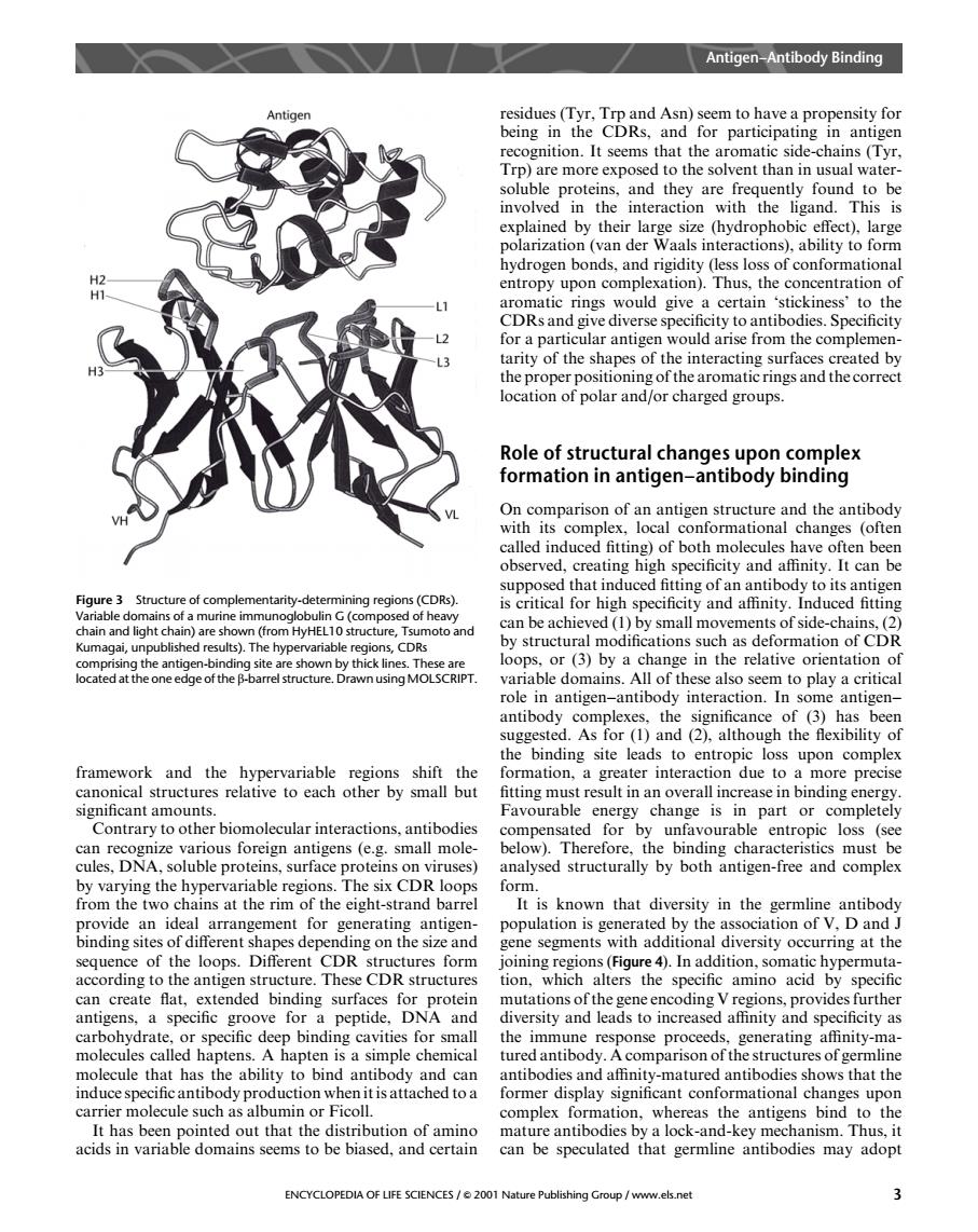
Antigen-Antibody Binding ition.It see T Trp)are more exposed to the solvent than in usual water solble and they found tob nvo gand. Sdogenboads,indngdryes1osofconormatiomal entropy upon complexation).Thus,the concentration o toRairdmeedongeacetainsticekine for a particular antigen would arise from the compleme tarity of the shapes of the interacting surfaces created by Role of structural changes upon complex formation in antigen-antibody binding od called induced fitting)of both molecules have often been observed,creating high specifcity and atfinity.It can be Figure 3 Structure of complementarity-detemining supposed thatn ats ofside chair by structural modifications such as deformation of CDR g the an ing site are shown by thickines. ated at the one ntibody int antibody co mplexes,the significance of (3)has been suggested.As for (1)and (2).although the flexibility of the binding site leads to entropic loss upon complex imore precis significant amounts. Favourable energy change is in part or completel Contrary to other biomolecular interactions.antibodies compensated for by unfavourable entropic loss (see structurally by be from the two chains at the rim of the eisht-strand barre It is known that diversity in the germline antibody a品adew器e population is generated by the association of V,D and J binding sites of different shapes depe segments with additional diversity occurring at the DR ferent The CDR can create flat,extended binding surfaces for mutations ofthe g antigens,a specific groove for a peptide,DNA and diversity and leads to increased affinity and specificity as generating athnity-ma chemica induce production to carrier molecule such as albumin or Ficoll. complex formation,whereas the antigens bind to the mature antibodies by antib ENCYCLOPEDIA OF LIFE SCIENCES/2001 Nature Publishing Group/www.els.net 3 framework and the hypervariable regions shift the canonical structures relative to each other by small but significant amounts. Contrary to other biomolecular interactions, antibodies can recognize various foreign antigens (e.g. small molecules, DNA, soluble proteins, surface proteins on viruses) by varying the hypervariable regions. The six CDR loops from the two chains at the rim of the eight-strand barrel provide an ideal arrangement for generating antigenbinding sites of different shapes depending on the size and sequence of the loops. Different CDR structures form according to the antigen structure. These CDR structures can create flat, extended binding surfaces for protein antigens, a specific groove for a peptide, DNA and carbohydrate, or specific deep binding cavities for small molecules called haptens. A hapten is a simple chemical molecule that has the ability to bind antibody and can induce specific antibody production when it is attached to a carrier molecule such as albumin or Ficoll. It has been pointed out that the distribution of amino acids in variable domains seems to be biased, and certain residues (Tyr, Trp and Asn) seem to have a propensity for being in the CDRs, and for participating in antigen recognition. It seems that the aromatic side-chains (Tyr, Trp) are more exposed to the solvent than in usual watersoluble proteins, and they are frequently found to be involved in the interaction with the ligand. This is explained by their large size (hydrophobic effect), large polarization (van der Waals interactions), ability to form hydrogen bonds, and rigidity (less loss of conformational entropy upon complexation). Thus, the concentration of aromatic rings would give a certain ‘stickiness’ to the CDRs and give diverse specificity to antibodies. Specificity for a particular antigen would arise from the complementarity of the shapes of the interacting surfaces created by the proper positioning of the aromatic rings and the correct location of polar and/or charged groups. Role of structural changes upon complex formation in antigen–antibody binding On comparison of an antigen structure and the antibody with its complex, local conformational changes (often called induced fitting) of both molecules have often been observed, creating high specificity and affinity. It can be supposed that induced fitting of an antibody to its antigen is critical for high specificity and affinity. Induced fitting can be achieved (1) by small movements of side-chains, (2) by structural modifications such as deformation of CDR loops, or (3) by a change in the relative orientation of variable domains. All of these also seem to play a critical role in antigen–antibody interaction. In some antigen– antibody complexes, the significance of (3) has been suggested. As for (1) and (2), although the flexibility of the binding site leads to entropic loss upon complex formation, a greater interaction due to a more precise fitting must result in an overall increase in binding energy. Favourable energy change is in part or completely compensated for by unfavourable entropic loss (see below). Therefore, the binding characteristics must be analysed structurally by both antigen-free and complex form. It is known that diversity in the germline antibody population is generated by the association of V, D and J gene segments with additional diversity occurring at the joining regions (Figure 4). In addition, somatic hypermutation, which alters the specific amino acid by specific mutations of the gene encoding V regions, provides further diversity and leads to increased affinity and specificity as the immune response proceeds, generating affinity-matured antibody. A comparison of the structures of germline antibodies and affinity-matured antibodies shows that the former display significant conformational changes upon complex formation, whereas the antigens bind to the mature antibodies by a lock-and-key mechanism. Thus, it can be speculated that germline antibodies may adopt Figure 3 Structure of complementarity-determining regions (CDRs). Variable domains of a murine immunoglobulin G (composed of heavy chain and light chain) are shown (from HyHEL10 structure, Tsumoto and Kumagai, unpublished results). The hypervariable regions, CDRs comprising the antigen-binding site are shown by thick lines. These are located at the one edge of the b-barrel structure. Drawn using MOLSCRIPT. Antigen–Antibody Binding ENCYCLOPEDIA OF LIFE SCIENCES / & 2001 Nature Publishing Group / www.els.net 3