正在加载图片...

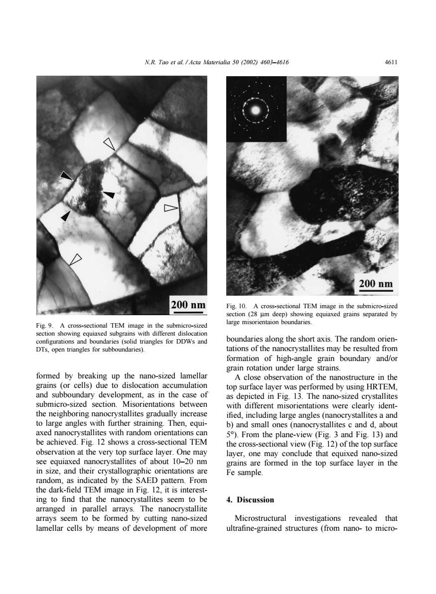
N.R.Tao et al.Acta Materialia 50 (2002)4603-4616 4611 200nm 200nm Fig.10.A cross-sectional TEM image in the submicro-sized section(28 um deep)showing equiaxed grains separated by Fig.9.A cross-sectional TEM image in the submicro-sized large misorientaion boundaries. section showing equiaxed subgrains with different dislocation configurations and boundaries(solid triangles for DDWs and boundaries along the short axis.The random orien- DTs,open triangles for subboundaries). tations of the nanocrystallites may be resulted from formation of high-angle grain boundary and/or grain rotation under large strains. formed by breaking up the nano-sized lamellar A close observation of the nanostructure in the grains (or cells)due to dislocation accumulation top surface layer was performed by using HRTEM, and subboundary development,as in the case of as depicted in Fig.13.The nano-sized crystallites submicro-sized section.Misorientations between with different misorientations were clearly ident- the neighboring nanocrystallites gradually increase ified,including large angles(nanocrystallites a and to large angles with further straining.Then,equi- b)and small ones (nanocrystallites c and d,about axed nanocrystallites with random orientations can 5).From the plane-view (Fig.3 and Fig.13)and be achieved.Fig.12 shows a cross-sectional TEM the cross-sectional view (Fig.12)of the top surface observation at the very top surface layer.One may layer,one may conclude that equixed nano-sized see equiaxed nanocrystallites of about 10-20 nm grains are formed in the top surface layer in the in size,and their crystallographic orientations are Fe sample. random,as indicated by the SAED pattern.From the dark-field TEM image in Fig.12,it is interest- ing to find that the nanocrystallites seem to be 4.Discussion arranged in parallel arrays.The nanocrystallite arrays seem to be formed by cutting nano-sized Microstructural investigations revealed that lamellar cells by means of development of more ultrafine-grained structures (from nano-to micro-N.R. Tao et al. / Acta Materialia 50 (2002) 4603–4616 4611 Fig. 9. A cross-sectional TEM image in the submicro-sized section showing equiaxed subgrains with different dislocation configurations and boundaries (solid triangles for DDWs and DTs, open triangles for subboundaries). formed by breaking up the nano-sized lamellar grains (or cells) due to dislocation accumulation and subboundary development, as in the case of submicro-sized section. Misorientations between the neighboring nanocrystallites gradually increase to large angles with further straining. Then, equiaxed nanocrystallites with random orientations can be achieved. Fig. 12 shows a cross-sectional TEM observation at the very top surface layer. One may see equiaxed nanocrystallites of about 10–20 nm in size, and their crystallographic orientations are random, as indicated by the SAED pattern. From the dark-field TEM image in Fig. 12, it is interesting to find that the nanocrystallites seem to be arranged in parallel arrays. The nanocrystallite arrays seem to be formed by cutting nano-sized lamellar cells by means of development of more Fig. 10. A cross-sectional TEM image in the submicro-sized section (28 µm deep) showing equiaxed grains separated by large misorientaion boundaries. boundaries along the short axis. The random orientations of the nanocrystallites may be resulted from formation of high-angle grain boundary and/or grain rotation under large strains. A close observation of the nanostructure in the top surface layer was performed by using HRTEM, as depicted in Fig. 13. The nano-sized crystallites with different misorientations were clearly identified, including large angles (nanocrystallites a and b) and small ones (nanocrystallites c and d, about 5°). From the plane-view (Fig. 3 and Fig. 13) and the cross-sectional view (Fig. 12) of the top surface layer, one may conclude that equixed nano-sized grains are formed in the top surface layer in the Fe sample. 4. Discussion Microstructural investigations revealed that ultrafine-grained structures (from nano- to micro-